Table of Contents
Most common reasons for a 6-Week Ultrasound Scan
The most common reasons for the 6 weeks and generally for early gestation age baby scans are:- Previous miscarriage.
- You had fertility treatment.
- Pelvic pain on one side
- Vaginal spotting or bleeding.
- You are unsure how far along you are in your pregnancy.
- You want a visual confirmation that you are pregnant after a positive pregnancy scan.
What should you see on a 6-week scan?
At 6 weeks gestation, you might be able to see:
- a black oval circle (black is fluid on ultrasound) which is the gestation sac
- A little white ring which is the yolk sac where the baby feeds from in the early weeks of pregnancy,
- the embryo (foetal pole)and
- possibly the heartbeat might be visible.
Depending on the individual pregnancy all the above however might not be visible during the scan.
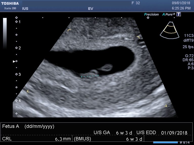
At 6 weeks, you won’t, in general, be able to see much detail of your baby. The ultrasound scan, however, should be able to confirm the gestation age by measuring either the gestation sac or the foetal pole if visible. Sometimes but not always you will be able to see the baby’s heartbeat.
Most importantly the sonographer who will do your private ultrasound in London will be able to check that your baby is in the right place i.e. in the endometrial cavity and that you do not have an ectopic pregnancy.
Ectopic pregnancy is when the fertilized egg attaches itself outside the uterus with the most common location being the fallopian tube on the side where you ovulated from.
Everyone obviously is different and sometimes a follow-up ultrasound in a week to 10 days later might be necessary to give you more information.
Your baby at 6 weeks
How many mm is a 6-week-old foetus?
At 6 weeks, your baby should measure approximately 5-9mms in length.
Can you see the baby at 6 weeks?
6 weeks into your pregnancy is also the earliest time that you might be able to see the foetal pole and the foetal heartbeat.
Can you see the baby heartbeat at the 6-week scan?
The foetal heartbeat looks like two parallel lines flickering, and it is not always visible at the 6-week scan. The literature suggests that the foetal heartbeat should be around 90-110 beats per minute, but we have seen slower heartbeats with positive pregnancy outcomes.
What else can you see?
The yolk sac, a ring shape bright circle might also be visible. The yolk sac is where your baby is feeding on at this early stage in pregnancy.
Sometimes only the gestation sac is visible with no foetal pole or yolk sac, and you might be asked to come back in a week to 10 days. In most cases, this is because you might be earlier in your pregnancy than you think.
What is the earliest you can have a pregnancy scan?
The 6-week scan is the most common gestation age that a private ultrasound is performed.
We do not recommend a scan before the 6 weeks gestation unless you are worried about a miscarriage or an ectopic pregnancy, as at 5 weeks gestation you will possibly see the endometrium being thickened and echo bright and possibly a gestation sac. A 5-week baby scan however might help to find the cause for any early pregnancy pain or bleeding.
If you are more than 6 weeks you may also want to read more about the 7-week scan and the 8+ week scan.
What happens at a 6-week scan?
It is more likely that at 6 weeks gestation age you will need to have a transvaginal or internal ultrasound scan instead of a transabdominal scan (through the abdomen). This is because it is a very early stage and everything is still very small. The transvaginal scan probe will be able to get closer to the endometrium and produce a better clearer image of the pregnancy insitu.
Feeling nervous about having an ultrasound scan so early in your pregnancy is normal. Try to stay calm and prepare yourself for what may happen. Bringing with you your partner or a close family member for extra support might be a good idea.
We do allow another family member present in the scanning room during the private ultrasound even during Covid-19 pandemic.
How many scans will I have during pregnancy through the NHS?
You will have at least two baby scans during your pregnancy provided by the NHS:
- a 12-week dating scan and
- a 20-week anomaly scan.
The 12-week scan will provide confirmation and dating for your pregnancy. The 20-week scan will provide information about your baby’s growth and development.
About Pregnancy Scans
A pregnancy ultrasound scan is the same as a ‘normal’ scan, but it is being used to evaluate the overall health of your baby instead of looking at other organs such as gallbladder for gallstones or kidney for kidney stones. So in pregnancy, ultrasound scans are being used to visualise the baby, the placenta, the uterus and cervix and your ovaries.
Pregnancy ultrasound scans or prenatal ultrasounds are very common and being carried in any stage of the pregnancy.
If you have any questions, or you want to know more about our private ultrasound in London please get in touch with us, and we will do our best to answer.
At our private ultrasound clinic, we offer pregnancy scans from as early as 5-6 weeks gestation in times to suit you as well as private scans for men and women.
Who interprets the results of the early pregnancy scan and how do I get them?
A Sonographer, a Health Care Professional specifically trained to perform and understand the ultrasound images, will most likely perform your exam and provide you with a digital ultrasound report that you can take it to your doctor or keep it for your records.
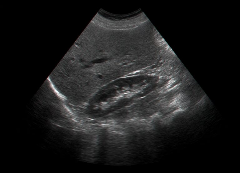
About Ultrasound
Diagnostic Medical ultrasound scan or medical sonography as otherwise known is a painless imaging technique utilising sound waves to produce internal images of the body.
It is called ultrasound as the sound frequency being used is at the region of 1 to 20MHz. The human ear can’t hear these frequencies.
The sound waves are produced by the transducer or the probe as most commonly known. As they travel through the body they bounce back to the transducer due to various sound transmissions differences in tissues. The returning echoes are picked up by the probe and a powerful computer analyses the echoes and creates the 2d image on the screen.
There are various kinds of ultrasound scans that can be performed and each looks at different organs of the body such as tendons, muscles, joints, blood vessels, liver, kidneys, uterus and ovaries to confirm or exclude possible pathology.
Unlike CT and MRI, pregnancy ultrasound does not use radiation and therefore is pregnancy-friendly. It is also live and is ideal for musculoskeletal exams to evaluate moving joints.
You can read more about the history of ultrasound.
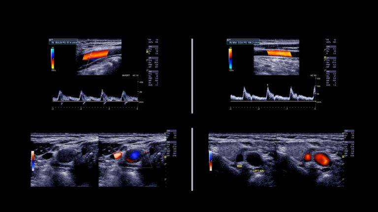
Other ultrasound scans related to pregnancy?
Some mothers to be will, unfortunately, get various complications during pregnancy such as high blood pressure, kidney infections and abnormal liver function tests. As ultrasound scans are pregnancy-friendly your doctor might refer you for an abdominal/liver scan or a kidney scan to check for anything that might explain your symptoms.
Although these ultrasound scans are not pregnancy scans, they are related to pregnancy.
In most cases, all the complications resolve after delivery, but like everything else related to your health and your baby’s health better safe than sorry.
What are ultrasound scans used for in pregnancy?
Depending on your stage of pregnancy, ultrasounds will be used to give you and your doctor or midwife answers about the health of your pregnancy.
First Trimester Ultrasounds
- Check that you are pregnant and that your baby has a heartbeat.
- Check if you have a singleton or twins
- Make sure that the pregnancy is not an ectopic located within the endometrial cavity and is not outside the womb such as in the fallopian tube.
- Look for the cause of any bleeding you might have.
- Date the pregnancy by measuring the crown-rump length of the foetal pole.
Second Trimester Ultrasounds
- Verify dates and growth
- Estimate the baby’s risk of Down’s syndrome by measuring the fluid at the back of your baby’s neck between about 10 weeks and 14 weeks
- Help with diagnostic tests by showing the position of the baby and placenta.
- Check your baby to see if all his organs are normal.
- Diagnose abnormalities
- Assess the amount of amniotic fluid and the location of the placenta.
- Evaluation of foetal well-being
Third-trimester Ultrasounds
- Make sure your baby is growing at the expected rate.
- Confirm if your baby is a boy or a girl.
Do you want to book your early pregnancy scan?
Private Pregnancy Scans
We offer the following complementary to the NHS private baby scans in our ultrasound clinic in London.


Glossary
Ultrasound: A medical imaging technique that uses high-frequency sound waves to create images of structures within the body, such as organs, tissues, or a fetus during pregnancy.
Transvaginal Ultrasound: A type of ultrasound scan where the ultrasound probe is inserted into the vagina to get clearer images of the pelvic organs or an early pregnancy.
Gestational Sac: The first ultrasound evidence of pregnancy, a fluid-filled structure surrounding the developing embryo in the uterus.
Yolk Sac: A small circular structure inside the gestational sac that provides nutrients to the developing embryo and is involved in the production of the embryo’s blood cells.
Fetal Pole: The first visual evidence of the developing embryo on an ultrasound scan, appearing as a thickening on the margin of the yolk sac, indicative of the early stages of fetal development.
Heartbeat: The rhythmic contraction and expansion of the heart, which can sometimes be detected as early as 6 weeks into pregnancy via ultrasound.
Crown-Rump Length (CRL): The measurement of the length of the fetus from the top of its head (crown) to the bottom of its torso (rump), used to estimate gestational age.
Gestational Age: The age of the pregnancy calculated from the first day of the last menstrual period (LMP) or via ultrasound measurements.
Viability: The ability of the fetus to survive outside the womb, which is generally considered from around 24 weeks of gestation. In the context of early pregnancy scans, “viability” refers to the potential of the pregnancy to continue under normal conditions.
Multiple Pregnancies: A pregnancy in which more than one fetus is developing, such as twins or triplets.
Ectopic Pregnancy: A potentially life-threatening condition where the embryo implants outside the uterine cavity, often in a fallopian tube.
Miscarriage: The spontaneous loss of a pregnancy before the fetus can survive outside the womb, typically defined as before 20 weeks of gestation.
Nuchal Translucency (NT): A collection of fluid under the skin at the back of a fetus’s neck, measured via ultrasound between 11 and 14 weeks of gestation as a marker for chromosomal abnormalities.
Chromosomal Abnormalities: Genetic disorders resulting from changes in the number or structure of chromosomes, such as Down syndrome.
Prenatal Care: Medical and nursing care recommended for women during pregnancy to ensure the best health outcomes for both mother and baby.
References:
NHS- Ultrasound Scans in Pregnancy
NHS – 12-week scan
ROOG – Guidance for antenatal screening and ultrasound in pregnancy in the evolving coronavirus pandemic
NICE – Antenatal care for uncomplicated pregnancies
BMUS – Guidelines for the safe use of diagnostic ultrasound equipment
Ultrasound in Obstetrics and Gynaecology – ISUOG Practice Guidelines: performance of first‐trimester fetal ultrasound scan
Healthline – Pregnancy Ultrasound
WHO – WHO recommendation on early ultrasound in pregnancy
Medically Reviewed by Tareq Ismail Pg(Dip), BSc (Hons)