Early detection of breast cancer is essential for improving mortality rates. Screening for this disease includes clinical breast exams, mammograms and ultrasound tests.
Expert groups generally suggest women perform regular breast self-exams (BSEs) to become familiar with their breasts' appearance and feel free to report any changes. BSEs cannot replace clinical breast examinations by healthcare providers, but they can help supplement screening efforts.
The Importance of Regular Breast Self-Examinations
Conducting regular breast self-exams is an integral part of women's overall healthcare regimen. It's a safe and straightforward way to detect any issues, such as lumps, so they can be treated promptly.
Women must know the warning signs of breast cancer before they are diagnosed, in order to receive timely treatment. According to the National Cancer Institute, those diagnosed with breast cancer in its earliest stages have the highest rates of survival and the best outcomes.
The American Cancer Society recommends that women perform monthly breast self-exams to detect cancer before it spreads. According to the National Breast Cancer Foundation, one out of eight women will develop breast cancer during their lifetime; thus, everyone should check their breasts regularly and report any changes to their doctor.
For a successful breast self-exam, it's essential that the patient has good control over her movements and is in an environment free from distractions.
Once the female is in a comfortable position to examine her breasts, she should use three middle fingers to feel for tissue. Make sure she covers the entire area without missing any breast tissue by applying different levels of pressure at different pressure points.
This type of breast exam, also referred to as a "palpable examination," can help detect cancer before it spreads and poses serious medical risks. Unfortunately, palpable examinations don't always detect small and early tumors, so getting a mammogram is recommended in addition.
It is essential that women perform a breast self-exam at the same time each month, whether they are still menstruating or have stopped. This will help them become familiar with when and how their exam should be performed each month.
It is essential that a breast exam not be conducted while wearing bras or other clothing, as this makes it difficult to feel the breast tissue. Furthermore, the exam should be performed while lying down so that all of your arms are free for examination of breast tissue.
The Importance of Regular Ultrasound Screening
Ultrasound screening is an imaging test that can detect certain types of breast cancer and other anomalies. It's particularly valuable as it may detect early signs of breast cancer, allowing treatment to begin sooner and giving the disease a better chance at being successfully treated.
Ultrasound is a safe and accurate imaging tool that can assist in diagnosing and treating many diseases and medical conditions. Additionally, it assists physicians when planning or performing specific procedures.
Can be performed in a doctor's office, clinic or hospital. Generally painless and without the use of ionizing radiation.
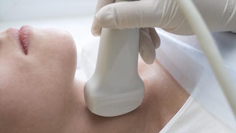
Ultrasound imaging involves a technologist (a person who works with sound waves) applying water-based gel on your skin in order to move a transducer over the area being studied. This helps lubricate the skin and conduct sound waves so they appear on computer screens.
Ultrasound can be performed on an outside surface or inside your body with a small probe. When performed externally, ultrasound is more precise since it can clearly show the organs beneath your skin.
A scan can take anywhere from 20 minutes to an hour, depending on how many parts of your body are being examined. A radiologist, or doctor trained to interpret radiology exams, will interpret the results and send them on to your healthcare provider.
Ultrasound is often the first imaging test a healthcare provider recommends for patients due to its cost-effectiveness and safety factor. Furthermore, ultrasound has more accuracy than X-rays or other tests when it comes to detecting cancerous or abnormal growths in the breast soft tissue.
Additionally, it does not use contrast agents (dyes) that are commonly used in other types of diagnostic imaging, making it safer for people who are allergic to these dyes.
This exam is performed on a table, with you lying flat. Depending on which part of your body needs checking, you may need to wear a gown.
The Importance of Regular Mammograms
As October marks National Breast Cancer Awareness Month, it is essential to educate ourselves and others about the significance of regular mammograms. These tests can assist in early detection of breast cancer and help reduce our overall risk for dying from this illness.
Mammograms are x-rays of the breast that can detect cancer in women without symptoms or signs. Additionally, mammograms can identify microcalcifications (tiny deposits of calcium) which could indicate cancer cells present.
Mammograms can be highly effective, but they also have their limitations. While they tend to be accurate, false-positive results may lead to unnecessary tests and biopsies. Furthermore, mammograms may cause distress for women diagnosed with cancer.
Most medical organizations recommend that women who are at average risk for developing breast cancer get annual mammograms. Screening helps detect cancer at an earlier stage when it can be more treatable and less likely to spread.
Mammograms should be done as soon as you reach the recommended age for them. If there has been a family history of breast cancer in your family, it's especially important to discuss this with your healthcare provider.
Another essential step is learning how to conduct a breast self-exam. This requires both visual and manual inspection of both breasts in both standing and reclining positions to look for any lumps or changes. It should be done monthly.
Studies have demonstrated that women who regularly perform BSE are more likely to detect tumors of 1 cm or smaller in size than those without this technique. Nonetheless, these findings have yet to be proven as a guarantee for improved survival rates from breast cancer.
Mammograms and self-exams are an excellent way for women to take control of their health and reduce the risk of breast cancer. These examinations help women become more aware of their breasts and how they typically look and feel, plus it enables them to report any changes immediately to their doctor if something seems abnormal.
The Importance of Regular Breast Biopsies
Regular breast self-examination and ultrasound screening are important methods for detecting the early signs of breast cancer. Both tests can be easily done, helping you reduce your risk for developing cancer by catching it at an earlier stage.
Self-exams should be performed monthly to detect any changes in the breast that could be concerning you or your doctor. The examination should include visual inspection and manual examination in various positions, such as standing and reclining.
In addition to performing a breast self-exam, you can also arrange an ultrasound appointment for a more comprehensive examination of your breasts. An ultrasound uses sound waves to look inside the breasts and can detect many types of abnormalities without using radiation like mammograms do. This test is much safer than performing self-exams as no radiation is used during its execution.
Ultrasound can detect how well blood flows through your breast tissue and even detect small cysts filled with fluid or solid tumors. It may also reveal microcalcifications - calcium deposits that aren't visible on mammograms or physical examination of your breast - which cannot be detected through mammography or physical exam alone.
Biopsies are another type of screening that can help your doctor detect any areas of concern in your breast. They're usually performed when a lump is discovered during a physical breast exam or when a mammogram reveals an unusual area.
Biopsies are typically not painful or damaging to breast tissue and the area where the needle is inserted will be cleaned and a shot of medicine injected to numb the skin.
Once the shot has numbed the area, a needle is inserted and tissue sample taken. After taking this sample, a sterile bandage is applied over the site to stop bleeding and absorb any fluid that collects there.
Breast biopsies come in two varieties: fine-needle aspiration (FNA) and core needle biopsy. FNA is used for lumps that can be felt with a physical exam, while core needle biopsy may be necessary when an imaging system cannot locate the lump or it's hard to reach.


