Breast implants are a popular cosmetic surgery procedure that can enhance breast size and shape. However, like any medical device, breast implants carry potential risks and may require monitoring over time. Breast ultrasound is an important imaging tool for evaluating breast implants and identifying any issues that may arise. This article provides a comprehensive overview of how and why breast ultrasound is used to monitor breast implants.
Overview of Breast Implants
Breast augmentation is the most common cosmetic plastic surgery procedure performed in the United States. In 2020 alone, over 300,000 breast augmentation surgeries were conducted [1]. The two main types of breast implants are:
- Saline implants – Saline implants are filled with sterile saltwater. They have a silicone outer shell and are filled with saline solution during surgery. Saline implants can be adjusted during surgery to achieve the desired size. If a saline implant ruptures, the implant will deflate quickly as the saltwater leaks out.
- Silicone implants – Silicone implants are pre-filled with a viscous silicone gel. They also have an outer silicone shell. Silicone implants are generally considered to have a more natural look and feel compared to saline. If a silicone implant ruptures, the gel may remain within the implant shell or only slowly leak out.
The most common reasons women choose to have breast augmentation include [2]:
- Increasing breast size and fullness
- Improving breast symmetry if breasts are different sizes
- Restoring breast volume lost after pregnancy/breastfeeding
- Improving proportions and contour for a more aesthetically pleasing shape
Breast implants carry certain risks, including [3]:
- Capsular contracture – scar tissue tightening around the implant causing hardness/distortion
- Rupture – the implant envelope tears, deflating saline implants or leaking silicone gel
- Infection
- Implant displacement/shifting
- Changes in nipple/breast sensation
Therefore, regular monitoring of breast implants is recommended to check for any signs of complication.
The Role of Ultrasound in Breast Implant Surveillance
Breast ultrasound is a key imaging tool for evaluating breast implants and identifying potential problems. The main reasons ultrasound is used for implant monitoring include:
- Detecting silent ruptures – Ultrasound can identify tears or holes in the implant outer shell and leakage of saline or silicone gel within the breast tissue. Ruptures are often “silent” with no obvious symptoms.
- Evaluating implant integrity – High-resolution ultrasound allows detailed assessment of the implant shell, valve sites, and contents. Saline implants can be checked for valve competence and saline fill volume.
- Assessing capsular contracture – Ultrasound can detect fibrous scar tissue formation around the implant and measure the thickness/density which indicates severity.
- Checking for complications – Ultrasound can identify implant-related complications like fold Fluid or hematoma formation, bottoming out/displacement, and lymph node swelling.
- Screening for breast cancer – Ultrasound is more accurate than mammography for detecting breast masses or abnormalities in augmented breasts, especially in dense breast tissue.
- Avoiding unnecessary MRI exams – Ultrasound is a lower cost non-ionizing imaging alternative that can evaluate and monitor implants without MRI in many cases.
- Guiding interventional procedures – Ultrasound allows precise needle guidance for aspirations, biopsies or injections related to implants.
Compared to mammograms and MRI, breast ultrasound offers several advantages for implant monitoring [4]:
- Lower cost and no radiation exposure
- Widely available even at small imaging centers
- Non-contrast enhanced procedure
- Simultaneous detailed imaging of breast tissue and implants
- Allows dynamic evaluation during compression/release
- Not affected by implant material or type
When Should Breast Ultrasound be Performed for Implant Surveillance?
Medical organizations provide guidelines on when women with breast implants should undergo imaging evaluations:
- Post-operative checks – Ultrasound should be performed shortly after surgery and at follow up visits to establish a baseline and check for early complications like hematoma or fluid accumulation [5].
- 1-3 years after surgery – Routine surveillance ultrasound is recommended 1-3 years post-op and then every 2-3 years for life to screen for silent rupture [6].
- Symptomatic patients – Patients presenting with breast pain, lumps, swelling, distortion or other concerns should receive a prompt ultrasound exam.
- Screening timeline – Annual ultrasound exams may be warranted in some high risk patients, such as those with a history of capsular contracture or implant rupture [7].
- Uncertain mammogram findings – Ultrasound should be performed after mammograms reveal suspicious areas or uncertain readings in women with augmented breasts.
- MRI screening – Ultrasound may precede MRI to avoid unnecessary MRI exams if no abnormalities are found.
Women should discuss an appropriate ultrasound monitoring schedule with their surgeon based on individual risk factors and implant details. More frequent or additional imaging may be needed beyond standard recommendations in some cases.
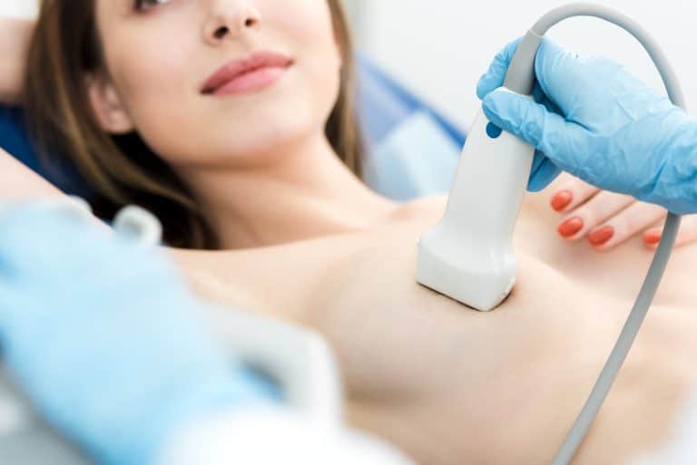
How is Breast Ultrasound Performed for Implant Evaluation?
Breast ultrasound exams for implant assessment involve detailed imaging of the breasts and implanted areas. The key components of a breast implant ultrasound include:
- High-frequency transducer – A linear array transducer with frequencies between 10-15 MHz or higher provides optimal resolution of breast implant structures. Lower frequencies (5-8 MHz) have deeper penetration for visualizing tissue behind implants [8].
- Bilateral evaluation – Both breasts are fully evaluated, including medial, lateral, inferior and posterior areas with comparison views of each side.
- Systematic scanning approach – Each area is scanned in transverse/axial and sagittal/longitudinal planes starting near the chest wall out to the nipple and entire breast. Radial scans may also be performed from the nipple outwards.
- Implant measurements – Implant dimensions, fill volumes for saline implants, shell thickness and implant pocket dimensions are measured.
- Compression technique – Moderate compression is applied during scanning to evaluate implant fold integrity, valve function, shell flexibility and identify crease fluid.
- Implant shell – The implant envelope is thoroughly examined for tears, holes, leaks, collapse, folds, valve site integrity and segmental contour irregularities.
- Peripheral assessment – The tissue surrounding the implants is evaluated for fluid collections, masses, lymph nodes or capsular complications.
- Adjunctive B-mode and color Doppler – Color flow imaging highlights ripples or color aliasing indicating rupture sites. Power/spectral Doppler quantifies suspicious internal fluid movements.
- Image documentation – Static images are recorded of all implant areas, along with video clips to allow comparison on serial exams.
Having a complete set of baseline images is important for following up potential issues over long-term implant surveillance.
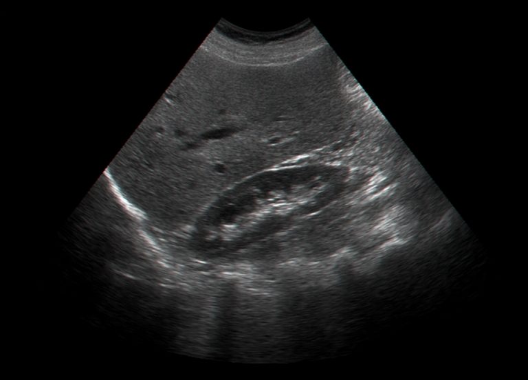
Indications of Breast Implant Rupture on Ultrasound
Detecting implant rupture is a major goal of surveillance ultrasound exams. There are various sonographic signs that may indicate an implant rupture:
Saline Implant Rupture
- Irregularity or collapse of implant outline, shape or folds
- Decreased implant volume – A saline implant should normally stay fully inflated
- Hyper-echoic lines extending from ruptured valve sites
- Direct visualization of a tear or hole in the implant shell
- Peri-implant fluid collection with internal echo debris
- New hyperechoic mass representing saline solution leakage
- Increased sound wave transmission and posterior enhancement
Silicone Implant Rupture
- Stepladder configuration – Collapsed upper half above a prevailingly filled lower half
- Gummy bear sign – Focal areas of adherent silicone putty against the implant shell
- Turbulent fluid motion on video compression
- Microcysts or microbubbles visualizing silicone droplets
- “Channel sign” – Fluid traversing silicone chambers through a ruptured barrier
- Snowstorm appearance – Echoic specks dispersed within the gel
- Milk of calcium sign – Layering hyperechoic calcifications
- “Strawberry pattern” – Hyperechoic dots settling in gravity-dependent locations
Other suspicious findings include:
- New peri-implant fluid collections or masses
- Enlarged or heterogeneous lymph nodes
- Increased vascularity around implants
- Skin thickening or Cooper’s ligament changes
Diagnosing Capsular Contracture with Breast Ultrasound
Another important purpose of breast ultrasound surveillance is detecting capsular contracture. This complication occurs when collagen fibers deposited around the implant during natural healing harden and contract. Signs of capsular contracture on ultrasound include [9]:
- Increased capsule thickness – Normally <2mm, >4mm indicates contracture
- Hyperechoic capsule appearance
- Straightened or angled posterior capsule contour
- Implant deformation – Teardrop shape, compression folds, diameter differences
- Higher echogenicity of implant contents
- Dilated or compressed peripheral veins
- Reduced compressibility and mobility during ultrasound
Capsular contracture is classified into 4 Baker grades based on physical exam findings. Ultrasound can determine the Baker grade which guides treatment:
- Grade I – Soft natural appearance, capsule <2mm
- Grade II – Minimal firmness, capsule 2-4mm
- Grade III – Noticeable distortion, >50% compressibility, capsule >4mm
- Grade IV – Severecontracture with painful rock-hard breasts, capsule >5mm
3D ultrasound provides even greater detail for evaluating breast capsule structure, thickness regularity and contracture severity.
Other Breast Implant Complications Detectable on Ultrasound
In addition to rupture and capsular contracture, ultrasound can also detect other potential breast implant complications:
- Seroma – peri-prosthetic fluid collection, anechoic on ultrasound
- Hematoma – blood accumulation near the implant, complex echogenic fluid
- Abscess – infected fluid collection with debris and increased vascularity
- Bottoming out – inferior displacement or sagging of the implant within the pocket
- Malposition – rotation or lateral displacement of the implant
- Extracapsular implant – shell herniation through the fibrous capsule
- Lymphadenopathy – enlarged or abnormal lymph nodes near implant
- Delayed wound healing – skin thickening, inflammation and fluid at incision site
Many of these issues require surgical correction after being identified on ultrasound. Careful screening allows complications to be diagnosed in early stages before symptoms become severe.
Breast Cancer Detection in Augmented Breasts
Women with implants can still develop breast cancer that needs to be evaluated on imaging. However, mammography has limitations in augmented breasts, especially in dense glandular tissue. Ultrasound is often a more effective tool for detecting breast masses or abnormalities after augmentation.
Signs of concern on ultrasound include [10]:
- Solid hypoechoic masses with irregular or microlobulated margins
- Combined cystic-solid lesions with thick walls and internal septations
- Masses with increased peripheral or internal vascularity
- Enlarged lymph nodes with eccentric cortical thickening
- Architectural distortion of breast tissue or skin thickening
Targeted ultrasound allows clear visualization and characterization of masses obscured on mammogram. Biopsy under ultrasound guidance can obtain samples from suspicious areas to make a definitive breast cancer diagnosis when indicated.
Guiding Interventional Procedures Related to Breast Implants
Ultrasound is an excellent tool for guiding interventional procedures involving breast implants given its real-time visualization and lack of radiation. Some examples include:
- Breast cyst aspiration – draining peri-implant fluid using ultrasound guidance
- Abscess drainage – inserting a catheter into a walled-off infected fluid collection under ultrasound
- Seroma aspiration – sampling peri-implant fluid for analysis
- Fine needle biopsy – sampling suspicious masses or lymph nodes seen on ultrasound
- Pre-operative measurements – using ultrasound to plan surgical approach based on precise implant measurements
- Injections – instilling drugs into the implant capsule using sonographic targeting
- Hematoma drainage – relieving pressure and pain from blood collecting around an implant
Ultrasound enables safe, effective needle insertion and aspiration without damaging implants during these interventions.
Considerations for Breast Ultrasound in Implant Patients
Breast ultrasound for implant evaluation requires some unique considerations:
- Higher frequency transducers optimize resolution but have limited penetration. Lower frequencies may be needed to image deeper structures behind or beneath implants.
- Gel-filled standoff pads are often required to prevent implant compression and allow complete visualization.
- Supine, upright weightbearing, and ipsilateral decubitus positions facilitate assessing implant stability, folds and leakage.
- Dynamic scans during compression evaluate shell integrity, fluid movements, and implant mobility within surrounding tissues.
- Knowing implant dimensions, material, fill volume and manufacturer helps tailor the exam and interpret findings.
- Images should be obtained for medial, lateral and posterior surfaces of each implant, requiring maneuvering probe positions.
- Follow up scans should replicate the same patient positioning, techniques and implant orientations to compare with baselines.
With proper understanding of implanted breast anatomy and optimized scanning protocols, radiologists can obtain the most accurate ultrasound evaluations.
Conclusion
Breast ultrasound imaging plays an integral role in the monitoring and surveillance of breast implants. It is the most effective way to screen for asymptomatic rupture, capsular contracture, and other complications. Ultrasound should be performed periodically starting 1-3 years after surgery and any time patients have breast concerns.
Compared to mammography and MRI, ultrasound provides a lower-cost and radiation-free method for evaluating augmented breasts without contrast agents. Ultrasound also enables image-guided aspirations, biopsies and injections involving implants when needed. With standardized exam techniques and recognition of sonographic findings, radiologists can provide optimal assessment of breast implants throughout a patient’s lifetime. Regular ultrasound surveillance allows early detection of silent problems and reduces future risks related to implants.
References
[1] American Society of Plastic Surgeons. 2020 Plastic Surgery Statistics Report. https://www.plasticsurgery.org/documents/News/Statistics/2020/plastic-surgery-statistics-full-report-2020.pdf
[2] Young VL. Breast Implants. In: StatPearls. StatPearls Publishing; 2022. https://www.ncbi.nlm.nih.gov/books/NBK448191/
[3] US Food and Drug Administration. Risks and Complications of Breast Implants. https://www.fda.gov/medical-devices/breast-implants/risks-complications-breast-implants
[4] Berg WA, Birdwell RL, Gombos EC. Diagnostic Imaging: Breast. Amirsys Publishing; 2004.
[5] American Society of Plastic Surgeons. Evidence-Based Clinical Practice Guideline: Breast Augmentation. https://www.plasticsurgery.org/Documents/Health-Policy/Guidelines/guideline-breast-augmentation.pdf
[6] Holmich LR, Fritzer N, Kjøller K, et al. The diagnosis of silicone breast implant rupture: clinical findings compared with findings at magnetic resonance imaging. Ann Plast Surg. 2005;54(6):583-589.
[7] Canadian Agency for Drugs and Technologies in Health. Optimal Imaging Strategies for the Detection and Surveillance of Breast Implant Associated Anaplastic Large Cell Lymphoma. 2018.
[8] Berg WA. Sonographic Evaluation to Detect Breast Implant Rupture. AJR Am J Roentgenol. 2009;192(6):1659-1660.
[9] Gorczyca DP, DeBruhl ND, Ahn CY, Shaw WW, Bassett LW. Silicone breast implant rupture presentation and diagnosis at MRI. Radiology. 1995 Apr;195(1):207-11.
[10] American College of Radiology. ACR Appropriateness Criteria – Silicone Breast Implant Surveillance. https://acsearch.acr.org/docs/3094102/Narrative/