Ultrasound imaging, also known as sonography or diagnostic medical sonography, has become an indispensable tool in modern medicine. Ultrasound uses high-frequency sound waves, above the range of human hearing, to produce dynamic visual images of organs, tissues, and blood flow inside the body.
The technology behind ultrasound is remarkably simple – it consists of a transducer probe which emits and receives sound waves as they bounce off tissues and structures. The probe is connected to a computer that processes the signals into 2D cross-sectional images in real-time. Ultrasound exams are completely safe, non-invasive, and painless.
Unlike techniques like X-rays, CT scans, or MRIs, ultrasound does not expose patients to ionizing radiation. It is also more portable and accessible than other imaging modalities. Ultrasound has a wide range of applications across medical specialties. It is particularly useful for observing soft body tissues that do not image well on X-rays.
Top 9 Uses of Ultrasound
1. Obstetrics
One of the most common uses of ultrasound is during pregnancy. Obstetric sonography is used to monitor fetal development, detect abnormalities, or confirm pregnancy health and viability. Ultrasound can provide views of the fetus, placenta, amniotic fluid, and uterus. It is typically performed at regular intervals during pregnancy to track fetal growth and position.
3D and 4D ultrasound can even produce realistic moving images of the fetus. Doppler ultrasound is used to examine blood flow in the umbilical cord and uterus. Ultrasound is a crucial obstetric tool, providing valuable information about fetal health and allowing early detection of potential issues.
2. Gynecology
In gynecology, ultrasound is routinely used to examine the female pelvic organs including the uterus, ovaries, cervix, and vagina. It helps identify uterine fibroids, ovarian cysts, endometriosis, or pelvic masses. Sonography is often the first imaging test for female reproductive issues.
Transvaginal ultrasound involves inserting a probe into the vagina to get closer images of the uterus and ovaries. This technique is used for detailed evaluation when pelvic examination is inconclusive.
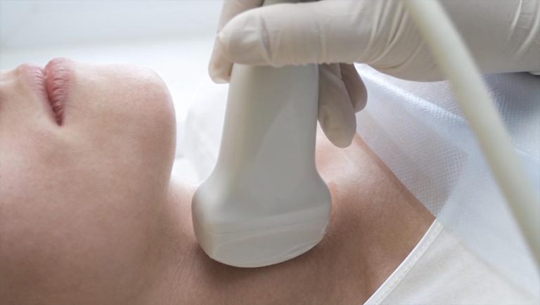
3. Cardiology
Echocardiography, a specialized type of ultrasound, is extensively used in cardiology. It produces images of the heart including chambers, valves, walls, and blood vessels. Echocardiogram can detect cardiomyopathies, abnormal anatomy, valve problems, or infections of the heart.
Stress echocardiography tracks heart function under exertion. Transesophageal echocardiogram (TEE) uses a probe inserted down the esophagus to get clear images. Echocardiography is affordable, portable, and plays a major role in diagnosing cardiac diseases.
4. Vascular
Ultrasound is often the first line of diagnosis for issues with blood vessels and circulation. Doppler ultrasound evaluates blood flow in arteries and veins, helping identify blockages or blood clots. This is useful in conditions like deep vein thrombosis, peripheral vascular disease, or aneurysms.
Carotid ultrasound specifically looks at the carotid arteries to check plaque buildup and supply to the brain. Arterial and venous mapping with ultrasound helps guide surgeries and placements of catheters or stents.
5. Musculoskeletal
MSK ultrasound is frequently used by orthopedists, rheumatologists, sports medicine specialists, and physical therapists. It can assess joint structures, muscles, tendons, and soft tissues in areas like the shoulder, knee, hips, elbows, wrists or ankles.
Ultrasound accurately detects muscle tears, damaged tendons, ligament sprains, ganglion cysts, or soft tissue masses. Ultrasound guidance assists procedures like joint aspiration or injections into tendon sheaths. Compared to MRI, ultrasound is a lower cost option for musculoskeletal imaging.
6. Ophthalmology
Eye ultrasound or ocular ultrasonography provides key anatomical information about the eye and orbit. It is useful when opacities in the eye obstruct other imaging techniques. Ultrasound can detect retinal detachment, intraocular tumors, or abnormalities in the lens, vitreous humor, and optic nerve.
Ultrasound biomicroscopy (UBM) utilizes high frequency probes to visualize anterior segment eye structures. Ophthalmic ultrasound is a valuable supplement to eye exams and optical tests like OCT or fundoscopy.
7. Breast
Breast ultrasound is routinely used alongside mammography to distinguish between solid masses and benign cysts. It helps determine whether a concerning lesion requires biopsy. Ultrasound aids guided biopsy collection and needle localization in breasts.
Supplemental screening ultrasound is recommended for women with dense breast tissue, implants or higher risk for breast cancer. Ultrasound elastography helps gauge tissue stiffness to predict malignancy. Portable ultrasound machines facilitate imaging in remote settings lacking mammography access.
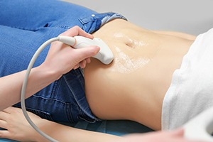
8. Abdominal
Abdominal ultrasound provides detailed views of the digestive organs and structures, including the liver, gallbladder, pancreas, kidneys, spleen, and abdominal aorta. It is used to identify gallstones, kidney stones, tumors, cysts, or obstructions in the abdominal cavity.
Abdominal ultrasound is often performed as follow-up to abnormalities or elevated liver enzymes found in blood tests. It also guides biopsies and drainages of abdominal fluid collections. Compared to CT, ultrasound does not use radiation and is preferred for pediatric patients.
9. Procedural Guidance
Real-time ultrasound visualization steers needles, catheters and probes during minimally invasive clinical procedures. Ultrasound guidance improves safety and precision for procedures like biopsies, aspirations, drainages, and catheter placements.
Breast, thyroid, liver, kidney and lymph node biopsies routinely rely on ultrasound guidance to advance the needle to the correct location. Vascular access procedures also greatly benefit from the accuracy of ultrasound visualization to avoid complications.
Recent Advances in Ultrasound Technology
Ultrasound technology has progressed tremendously in the last decade, with improvements in image quality, portability, advanced applications and integration with other modalities.
- Higher frequency broadband probes provide more detailed resolution down to microscopic levels.
- 3D and 4D ultrasound generate multiplanar and rendered volumetric views for enhanced analysis.
- Elastography and contrast-enhanced ultrasound have expanded ultrasound capabilities.
- Miniaturized and portable units allow ultrasound at point-of-care and bedside.
- Fusion imaging integrates ultrasound with other modalities like CT or MRI for combined benefits.
- Artificial intelligence is being applied to ultrasound for automated analysis and improved workflow.
Conclusion
Medical ultrasound has become an indispensable imaging tool integral to modern medical diagnosis and treatment across specialties. It provides real-time, high-resolution views of anatomical structures and organs in a versatile, non-invasive, radiation-free, and relatively low-cost manner.
With recent technological advances making the modality even more powerful and accessible, ultrasound will continue to expand into new applications and replace more costly or harmful imaging in many appropriate situations. Ultrasound’s utility and safety profile make it well-suited for use in outpatient clinics, at bedside, or in remote areas.