
Private Shoulder Ultrasound Scan in London
The tendons are watched while your arm moves — something no still image can show. Findings explained on screen as we scan, by the sonographer scanning you. No GP referral required.
- 30-minute appointment — unhurried, where most clinics allow 20
- HCPC-registered sonographers with senior NHS roles
- Findings at the scan; written report usually within two hours
- No GP referral required — self-refer today
- Report and images included — no hidden fees
CQC-registered clinic · Provider ID 1-2775844974 · HCPC-registered sonographers
What is a shoulder ultrasound scan?
A shoulder ultrasound uses high-resolution sound waves to examine the soft tissues around the shoulder joint: the four rotator cuff tendons, the bursa that cushions them, the biceps tendon at the front, and the acromioclavicular joint on top. There is no radiation and no injection.
Its defining advantage is movement. The scan happens while you lift, rotate and lower the arm, so a tendon can be watched sliding under the bone and a bursa seen catching as it does. A shoulder that looks unremarkable at rest can reveal its problem the moment it is put under load — and that is information a static image cannot produce. For rotator cuff tears in experienced hands, ultrasound performs comparably to MRI.
Why might I need a shoulder scan?
Most shoulder pain that has outlasted a few weeks comes from a small number of structures, and they are all within reach of ultrasound. The pattern usually points the way: pain lifting the arm out to the side suggests the rotator cuff and the bursa above it; pain at the front worse turning the palm up suggests the biceps tendon; pain on top, tender to press, suggests the AC joint.
Sudden weakness after a fall or a heavy lift is different again — that raises the question of a tear, and the answer changes what happens next. A scan turns “my shoulder hurts” into a named structure with a measurement beside it.
- Pain lifting the arm overhead or out to the side
- Night pain when lying on that shoulder
- Sudden weakness after a fall or heavy lift
- Front-of-shoulder pain, worse carrying or turning the palm up
- Pain on top of the shoulder, tender to press
- A shoulder that has not settled after weeks of physiotherapy
- All four rotator cuff tendons — tendinopathy, partial and full-thickness tears
- Subacromial bursa — bursitis and impingement, watched as you lift
- Biceps tendon — including dislocation out of its groove
- Acromioclavicular joint
- Calcific deposits within the tendons
- The other shoulder, for comparison where it helps
A visibly deformed shoulder after injury, complete inability to move the arm following trauma, or a hot swollen shoulder with fever needs same-day assessment — A&E or NHS 111. Pain spreading into the chest or jaw with breathlessness should be treated as a possible cardiac emergency: call 999. The scan can wait; these cannot.
Is this scan suitable for me?
A shoulder ultrasound is a good fit if any of these sound like you:
- ✓Your pain is worse lifting, reaching or sleeping on that side
- ✓You have lost strength in the arm since an injury
- ✓Physiotherapy has stalled and you want to know what is actually there
- ✓A previous scan or X-ray did not explain your symptoms
- ✓You want a tendon assessment before deciding on an injection or surgery
- ✓You would rather avoid radiation and start with the least invasive test
And when it is not the right booking
Pins and needles down the arm — that usually comes from the neck, not the shoulder, and needs a different assessment through your GP.
Suspected fracture or arthritis — bone is an X-ray question. We do not offer X-ray.
Suspected labral tear or instability — the labrum sits inside the joint and needs MR arthrography. We will tell you that before you book, not after.
If a shoulder ultrasound is not the right test for you, we will say so before you book — not after.
What happens during the examination?
- 1
Arrive — no preparation neededNothing to fast for and no full bladder. Wear something that leaves the shoulder easy to reach. The appointment is 30 minutes end to end.
- 2
Your history firstWe ask where it hurts, what movement provokes it and what you have already tried. That shapes which structures get the closest attention.
- 3
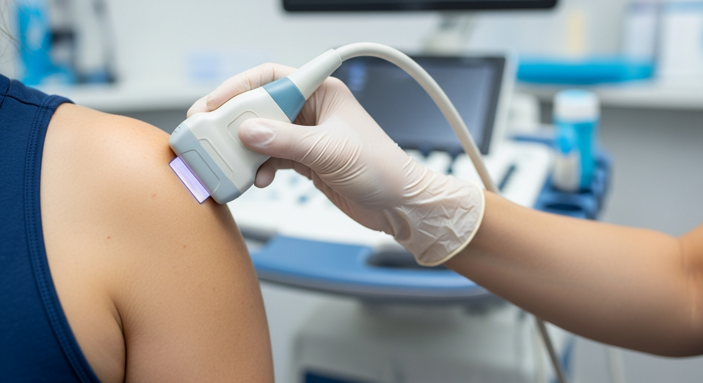
Scanning at restYou sit while warm gel and a high-resolution linear transducer are moved over the cuff, bursa, biceps tendon and AC joint.
- 4
Scanning under loadWe ask you to lift, rotate and lower the arm so the tendons are watched moving. This is where impingement and subtle tears declare themselves.
- 5
Comparison where it helpsThe other shoulder is often scanned too — tendon appearances vary between people, and your own other side is the best reference.
- 6
Findings explained on screenYou see what we see, described in plain terms, before you leave the room. The written report follows, usually within two hours.
Understanding the shoulder
Three structures explain most shoulder pain. Knowing which one is involved is what changes the treatment.
Rotator cuff
Four tendons wrap the top of the humerus and hold it centred in the socket while the larger muscles move the arm. Degeneration, impingement and tearing here account for the majority of shoulder pain that persists beyond a few weeks.
Subacromial bursa
A thin fluid-filled cushion between cuff and bone. When the space narrows, the bursa is compressed on every lift — producing the classic painful arc that eases once the arm is above the head.
Biceps tendon
The long head runs in a shallow groove at the front of the humerus and is vulnerable where it turns into the joint. It can inflame, tear, or slip out of its groove entirely — visible only when the arm rotates.
What your report might say
A finding is not automatically your diagnosis. Cuff tears and degenerative change are common in people with no shoulder pain at all, and their prevalence rises with age. What makes a finding relevant is whether it sits where you hurt and fits your story — which is why the report is written to be read alongside an examination, not instead of one.
Find out what is actually there
A 30-minute assessment with the tendons watched under load, explained on screen as we scan, with your written report usually within two hours.
How we scan shoulders
Scanned by the person reporting it
The sonographer who scans you writes your report. Nothing is handed to a remote reader who never met you or your shoulder.
Dynamic assessment as standard
Every shoulder is examined moving, not just at rest. Impingement and subtle tears are frequently invisible on a static image.
Both shoulders when it helps
Tendon appearance varies between people. Comparing with your other side is often what makes a borderline finding interpretable.
Honest routing
If your question needs MRI, X-ray or a surgical opinion, the report says so. We would rather send you to the right test than sell you ours.
Shoulder ultrasound — your questions answered
Is ultrasound as good as MRI for the shoulder?
Do I need a GP referral?
How long does the appointment take?
When will I get my results?
Can a scan diagnose frozen shoulder?
Does the scan hurt?
My shoulder hurts most at night. Is that a bad sign?
Can you inject the shoulder while I am there?
Should I have an X-ray as well?
What if the scan is normal but I am still in pain?
Other MSK scans we offer
Tendons, ligaments, bursae and swellings around the knee, assessed under load.
See the scan →
ElbowElbow Ultrasound Scan
Tennis and golfer’s elbow, bursitis and the ulnar nerve in real time.
See the scan →
All MSKOther MSK Scans
Hip, ankle, foot, hand and wrist — the full musculoskeletal range.
See scans →
Further reading: persistent shoulder pain — when to get an ultrasound and understanding your MSK report.
Where can you get a shoulder ultrasound in London?
Our clinic is at 5a Lucerne Mews, Kensington, London W8 4ED — a quiet mews three minutes on foot from Notting Hill Gate, serving Kensington, Notting Hill, Holland Park, Bayswater and central London.
Short-notice appointments are often available, including for people who have been given a long wait elsewhere. Call 0203 051 6506 for today’s availability.
Getting here
By tube: Notting Hill Gate — three minutes’ walk.
By bus: Routes along Notting Hill Gate and Kensington Church Street stop nearby.
By car: On-street parking nearby — full options in directions & parking.
Tendons watched while they work
A full shoulder assessment — cuff, bursa, biceps tendon and AC joint — with the arm moved through range, explained on screen as we scan. Self-refer today, no GP letter required. Short-notice appointments often available.
Kensington, London W8 4ED
3 mins from Notting Hill Gate
Written and clinically reviewed by the HCPC-registered sonographers who perform this scan at IUS London — a CQC-registered diagnostic ultrasound clinic (Provider ID 1-2775844974). Your own findings are explained to you at the scan and set out in your report.
Author: Yianni Kiromitis, Senior Sonographer, HCPC RA38415 — over 20 years’ experience in NHS and private ultrasound · Reviewed 10 August 2026