
Carotid Artery Ultrasound Scan in London
The two arteries in your neck supply most of your brain. We examine, measure and quantify their health in 30 minutes — without radiation. Plaque found early is a prevention plan, not a sentence.
- 30-minute unhurried examination — most clinics allow 20
- HCPC-registered sonographers with senior NHS roles
- Findings at the scan; written report usually within two hours
- No GP referral required — self-refer today
- Report and images included — same-day appointments often available
CQC-registered clinic · Provider ID 1-2775844974 · HCPC-registered sonographers
What is a carotid artery ultrasound?
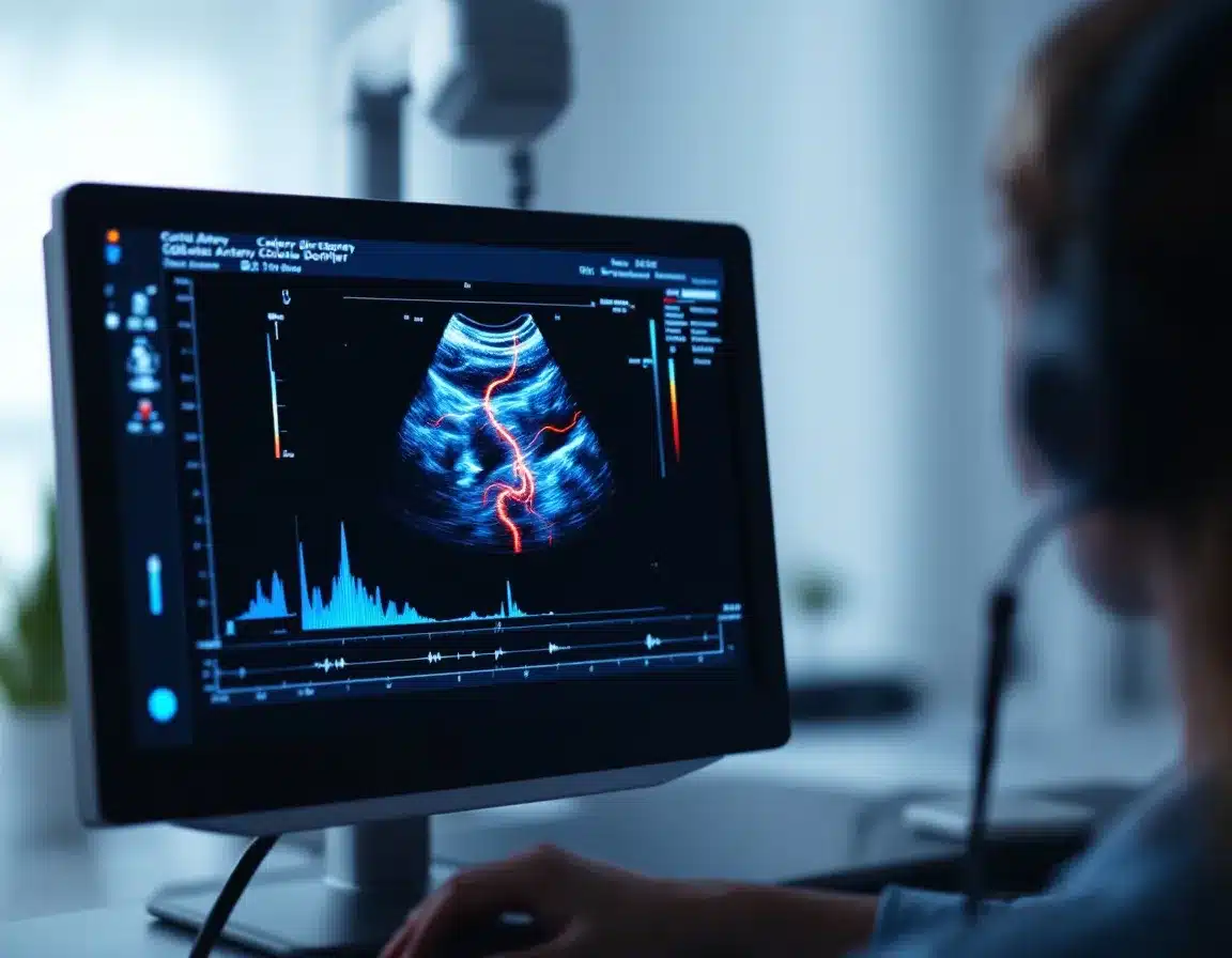
A carotid ultrasound is a painless Doppler examination of the two main arteries in your neck — the vessels that supply most of your brain. Using sound waves and colour flow mapping, it shows the artery walls directly, identifies any plaque, and measures the speed of your blood flow, which is how any narrowing is quantified precisely rather than guessed at. No needles, no radiation, no preparation — gel, a probe on your neck, and your own circulation on the screen in front of you.
Why might I need a carotid artery scan?
A strong family history of stroke often sits quietly on your mind. Perhaps you have known cardiovascular risk factors — high cholesterol, raised blood pressure, diabetes, or a smoking history — that make you want a direct, objective look at the vessels themselves.
Many patients book after a relative's stroke; others include it in a considered, proactive health review, or come because their GP suggested carotid imaging would usefully inform a prevention plan. A carotid scan replaces uncertainty with hard numbers. Our guide to carotid screening and stroke risk covers who benefits most.
Sudden face drooping, arm weakness, speech difficulty, vision loss or one-sided numbness — even if it passes within minutes — warrants a 999 call, not a scan booking. The NHS runs a same-day assessment pathway for exactly this, and it is excellent. The scan can wait; the brain cannot.
- High cholesterol
- High blood pressure
- Type 2 diabetes
- Smoking history
- Family history of stroke
- Plaque presence & type
- Stenosis severity
- Flow velocities
- Vessel wall changes
- Vertebral artery flow
Is this scan suitable for me?
A carotid ultrasound is a good fit if any of these sound like you:
- ✓A parent or sibling has had a stroke or TIA, and it sits on your mind
- ✓You have high cholesterol, high blood pressure, diabetes or a smoking history
- ✓Your GP suggested carotid imaging would inform your prevention plan
- ✓You want a measured cardiovascular baseline as part of a proactive review
And when it is not the right booking
Recent TIA or stroke symptoms — the NHS rapid-access pathway is faster, more complete, and free: that comes first, always.
Dizziness as your only symptom — carotid narrowing is rarely the cause; we will say so honestly, and the scan can still document a baseline if you want one.
If a carotid scan is not the right test for you, we will say so before you book — not after.
What does a carotid ultrasound actually show?
With colour and spectral Doppler we map both carotid systems in their entirety — the common carotid, the bifurcation where plaque most commonly forms, and the internal and external branches. We assess the artery walls directly, characterise any plaque, and measure your blood-flow velocities: when an artery narrows, blood accelerates through the restriction, and those velocities are how narrowing is precisely quantified against established clinical criteria.
- 1
Common carotid arteryBilateral assessment of the main vessel from the collarbone to the bifurcation, including wall changes.
- 2
Bifurcation & plaque assessmentThe carotid bulb examined in detail — any plaque characterised by location, size, echogenicity and surface.
- 3
Internal carotid — stenosis quantificationSpectral Doppler velocities grade any narrowing against established criteria — the numbers that change management.
- 4
Vertebral artery flowFlow direction and waveform in both vertebral arteries complete the picture.
Understanding the carotid artery system
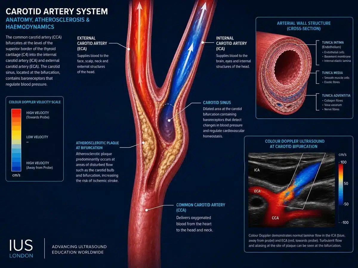
The carotid arteries are the brain's primary blood supply. Understanding the anatomy explains why the bifurcation is the vulnerable site — and why measured flow tells the truth.
Why the bifurcation?
Turbulent flow where the artery divides creates shear stress on the vessel wall — the main reason plaque forms at this precise anatomical site.
Wall changes come first
Early thickening of the artery wall is a quantifiable marker of vascular ageing — often measurable years before any significant narrowing develops.
Doppler velocimetry
Faster flow through a narrowing is measurable to the centimetre-per-second — which is how a stenosis is graded objectively rather than estimated by eye.
Understanding your results and next steps
We keep the tone calm and educational — because the facts support calm. Plaque is a very common sign of ageing vessels; finding some is the norm from midlife onwards. What matters is how much, where, and what it does to your flow — and every pathway is explained before you leave, with your report structured for your GP to act on.
How narrowing is graded
Blood accelerating through a narrowed segment is measured directly, and the velocity bands map to stenosis grades used across UK vascular practice:
| Finding | What it means |
|---|---|
| Normal velocities | Baseline documented |
| Moderate narrowing | GP prevention plan |
| Severe narrowing | Vascular referral |
| No flow detected | Urgent referral |
Graded against established UK consensus velocity criteria; your report states the exact measurements.
Thirty unhurried minutes between you and a measured answer — short-notice appointments often available.
The IUS London standard of care
A carotid scan's best outcome isn't a clean bill of health — it's a prevention plan started ten years early. We show you exactly what we're measuring and put every number in context.
HCPC-registered sonographers
Every scan performed by an HCPC-registered sonographer — clinicians who also hold senior NHS imaging roles.
Colour & spectral Doppler
High-resolution linear transducers with full Doppler capability — the image quality accurate grading depends on.
GP-ready report in ~2 hours
Your report carries the exact measurements and grading your GP needs — written to be acted on, usually within two hours.
Findings explained at the scan
You leave understanding what was found and what happens next — not waiting anxiously for a letter.
Why do patients choose IUS London?
Rated 4.9 across 476 Google reviews:
“Yianni was very helpful, knowledgeable and put me very much at ease. I will be going back and very much recommend.”
— Google review
“The staff were professional and made me feel comfortable. Highly recommend it!”
— Google review
“Amazing service. If you are looking for a clinic … this is the place to go.”
— Google review
Carotid scans — your questions answered
How much does a carotid artery scan cost?
Is finding plaque bad news?
Can this scan predict a stroke?
I've had a TIA — should I book this?
Do I need a GP referral?
How do I prepare?
Can this scan diagnose the cause of dizziness?
Is this the same as a "stroke screening test"?
When will I receive my report?
Where is the clinic?
Other vascular ultrasound scans we offer
Further reading: carotid screening and stroke risk — who should be scanned? · Considering a broader review? The well-man and well-woman scans include the aorta, with carotid as a recommended add-on.
Where can you get a carotid ultrasound in London?
At IUS London — our CQC-registered clinic in Kensington, three minutes on foot from Notting Hill Gate station (Central, Circle and District lines).
5a Lucerne Mews, Kensington, London W8 4ED
30-minute appointments — short notice welcome
0203 051 6506 · Directions & parking
Getting here is the easy part
By tube: Notting Hill Gate — three minutes' walk.
By bus: Routes along Notting Hill Gate and Kensington Church Street stop nearby.
By car: On-street parking nearby — full options in directions & parking.
Measure the risk you can't feel
Both carotid arteries mapped, narrowing quantified, prevention informed. Self-refer today — no GP letter required. Short-notice appointments available — call for today's availability.
Kensington, London W8 4ED
3 mins from Notting Hill Gate
Written and clinically reviewed by the HCPC-registered sonographers who perform this scan at IUS London. Your own findings are explained to you at the scan and set out in your report. Performed by HCPC-registered sonographers at our CQC-registered clinic (Provider ID 1-2775844974).
Author: Yianni Kiromitis, Senior Sonographer, HCPC RA38415
Medically reviewed: 25 July 2026