
Thyroid and Neck Ultrasound in London
Expert assessment of your thyroid gland, cervical lymph nodes and salivary glands — findings explained while you watch, by the sonographer scanning you.
- 30-minute unhurried examination — most clinics allow 20
- HCPC-registered sonographers with senior NHS roles
- Findings at the scan; written report usually within two hours
- No GP referral required — self-refer today
- No preparation — eat, drink and take medication as normal
CQC-registered clinic · Provider ID 1-2775844974 · HCPC-registered sonographers
What is a thyroid and neck ultrasound?
A thyroid and neck ultrasound is a safe, radiation-free imaging examination that uses high-frequency sound waves to build a detailed, real-time picture of your thyroid gland, the lymph nodes of your neck and your salivary glands. It is the first-line imaging investigation for thyroid conditions and neck lumps, in line with NHS and British Thyroid Foundation guidance, because it resolves superficial structures in exceptional detail, images in real time, and can be repeated as often as monitoring requires.
The thyroid itself is a butterfly-shaped endocrine gland at the front of your neck, just below the Adam’s apple. It produces the hormones that regulate your metabolism, energy, heart rate and body temperature, and it can develop a range of conditions: nodules and cysts, enlargement (goitre), inflammation (thyroiditis) and, rarely, cancer. The same appointment also examines the cervical lymph node chains, the parotid and submandibular salivary glands, and — where enlarged — the parathyroid glands behind the thyroid.
One honest limit, stated up front: ultrasound shows the structure of your thyroid; it does not measure how well the gland is working. Structure and function together give the full picture, and the two investigations prompt each other — for how they pair, read our guide to thyroid ultrasound questions answered.
Why might I need a thyroid and neck scan?
Most people book this scan for one of three reasons: something they can feel — a lump or swelling in the neck, in front of the ear or under the jaw; something they can feel happening — difficulty swallowing, a sensation of something stuck in the throat, persistent hoarseness, or pressure in the neck; or something a blood test has raised — abnormal thyroid function results that now need a structural explanation.
Others come for surveillance of a known nodule, after thyroid surgery, with a family history of thyroid disease, or with the classic dysfunction picture — unexplained weight change, persistent fatigue, palpitations, temperature sensitivity — where the scan documents the gland while blood tests measure its output.
- Neck lump or swelling
- Difficulty swallowing
- Hoarseness or voice change
- Swelling by the ear or jaw
- Neck pain or tenderness
- Thyroid size, texture, blood flow
- Nodules and cysts
- Cervical lymph nodes
- Parotid & submandibular glands
- Surrounding neck structures
Noisy or laboured breathing, a neck that is swelling rapidly, or a lump growing week on week warrant a 999 call or A&E now, not a scan booking. The NHS runs same-day assessment for exactly this, and it is excellent. The scan can wait; your airway cannot. For urgent but non-emergency concerns, tell us when booking and we will prioritise your appointment.
Is this scan suitable for me?
A thyroid and neck ultrasound is a good fit if any of these sound like you:
- ✓A lump or swelling in your neck you can see or feel
- ✓Difficulty swallowing, or a persistent feeling of something stuck in the throat
- ✓Hoarseness or a voice change that has lasted more than a few weeks
- ✓Abnormal thyroid blood tests that now need a structural explanation
- ✓A known thyroid nodule due its surveillance scan
- ✓Swelling or pain in front of the ear or under the jaw, especially when eating
- ✓A family history of thyroid disease and you want a documented baseline
And when it is not the right booking
Your only question is whether your thyroid is overactive or underactive — ultrasound cannot answer that. Gland function is measured by a simple blood test (TSH, T4, T3), usually arranged through your GP, and that is the right first step. Where the bloods are abnormal, or something can be felt, the scan then explains the structure behind the numbers.
If a thyroid ultrasound is not the right test for you, we will say so before you book — not after.
What does the scan actually cover?
Zone by zone, not one glide. Both lobes, the isthmus, the region behind the trachea and the node chains down each side are covered as named zones rather than a single sweep, with angle and pressure varied deliberately at each. Small nodules tuck behind the trachea and under the strap muscles, and a quick pass simply does not reach them — the point of the systematic order is that nothing is left to chance. Sides are compared as we go: right lobe against left, central neck against lateral.
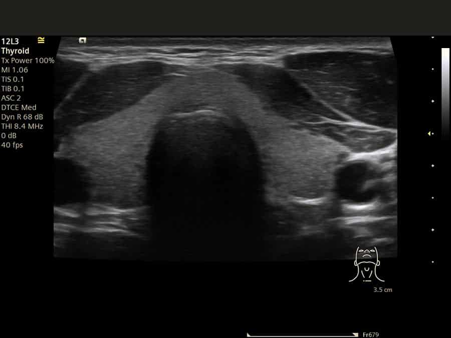
The thyroid gland. We measure the size and volume of each lobe, assess the texture and echogenicity of the tissue, and use colour Doppler to evaluate blood flow — the pattern that helps identify inflammation and autoimmune thyroid disease such as Hashimoto’s or Graves’.
Any nodule, characterised properly. Nodules are graded using TI-RADS, the standardised reporting system used across the NHS. Smooth borders, uniform internal structure and simple cysts point one way; irregular borders, microcalcifications, a taller-than-wide shape or increased internal blood flow point another, and may warrant fine needle aspiration for a definitive answer.
The lymph nodes, systematically. The cervical chains are assessed by level — levels I to VI — for size, shape, internal architecture and blood flow, distinguishing normal reactive nodes responding to infection from nodes that need further investigation.
Salivary glands and neighbours. The parotid and submandibular glands are examined for stones, inflammation, cysts and masses — the common culprits behind facial swelling or pain on eating — and enlarged parathyroid glands can sometimes be visualised in the work-up of calcium disorders.
- Ultrasound cannot diagnose cancer definitively — that requires a tissue sample (biopsy)
- It cannot measure thyroid function — that is a blood test
- Structures deep in the neck can be beyond its reach — where appropriate, we recommend the complementary investigation
- 1
BeforeNo preparation. Eat, drink and take medication as normal. Wear something with easy access to your neck, and leave necklaces at home.
- 2
DuringYou lie back with your neck slightly extended. Warm gel, a gentle transducer, several angles; you may be asked to swallow or turn your head. Completely painless.
- 3
Findings, liveYour sonographer explains what they are seeing as they scan and answers your questions in real time — no anxious wait.
- 4
AfterThe gel wipes off and you carry on with your day immediately. Findings are explained at the scan; your written report usually follows within two hours.
Understanding your thyroid
The gland, the hormones and the grading system — because a report you understand is worth more than a report you carry.
The feedback loop
The pituitary releases TSH to drive the thyroid; the thyroid answers with T4 and T3. When hormone levels fall, TSH rises — when they climb, TSH falls. That is why TSH is usually the first and most sensitive signal that something is off, long before symptoms make sense.
Structure meets function
The scan shows what your gland is; blood tests show what it does. Abnormal bloods send us looking for a structural cause; a structural finding sends you for function and antibody tests. Neither alone is the full picture — together they usually are.
TI-RADS, in plain language
Every nodule is scored on its appearance — shape, borders, content, calcification — using TI-RADS, the standardised system radiology teams use nationwide. Your next clinician reads the grade and knows exactly what was seen and what follow-up that appearance warrants.
Understanding your results and next steps
Worth holding onto: thyroid cancer is relatively rare and highly treatable, and the commonest type — papillary — carries an excellent prognosis with appropriate treatment. For the warning signs that do matter, read our guide to the symptoms of thyroid cancer.
Ready for a clear answer? £179, thirty unhurried minutes, findings explained as they appear.
The IUS London standard of care
An unhurried half hour
Thirty minutes booked for you alone — most clinics allow twenty. Enough time to scan the gland, the node chains and the salivary glands thoroughly, and to explain everything properly.
Tuned to the tissue, not the preset
Depth, gain and focus are re-optimised for each structure. A nodule that looks bland on default settings can reveal microcalcifications, irregular margins or increased blood flow once the image is tuned to it — and those are exactly the features that decide whether it is watched, sampled or left alone.
A report that moves fast
Findings are explained at the scan itself, and your written report usually follows within two hours — worded for your GP, endocrinologist or surgeon to act on.
Graded in the NHS’s language
Nodules are reported using TI-RADS, the standardised classification used across the NHS — so whoever sees you next knows exactly what was seen, without re-scanning.
Why do patients choose IUS London?
“I had a Thyroid and Neck ultrasound scan with Yianni … very knowledgeable and patient, taking the time to explain everything clearly. Really appreciate his professionalism and kindness — highly recommend.”
— Google review
“I had a thyroid and neck scan at IUS and had such a great experience … everything was explained in simple, easy-to-understand terms. I left feeling well looked after and so reassured. I highly recommend!”
— Google review
“Yianni was very helpful, knowledgeable and put me very much at ease. I will be going back and very much recommend.”
— Google review
Thyroid and neck scans — your questions answered
Do I need a GP referral for a thyroid ultrasound?
Do I need to prepare for the scan?
What happens during the appointment, and how long does it take?
When do I get my results?
Can an ultrasound diagnose thyroid cancer?
What happens if you find a thyroid nodule?
Will the scan show whether my thyroid is underactive or overactive?
Is the scan safe during pregnancy?
Are lymph nodes and salivary glands included?
How much does it cost, and are there hidden fees?
Other general ultrasound scans we offer
Soft-tissue lumps anywhere on the body — cysts, lipomas and lymph nodes named on screen, with honest answers on solid masses.
Explore this scan →
General ScansAbdominal UltrasoundLiver, gallbladder, pancreas, spleen, kidneys and aorta assessed in one unhurried examination.
Explore this scan →
General ScansKidneys, Ureters & Bladder (KUB)The renal tract end to end — stones, obstruction and bladder emptying with post-void residual.
Explore this scan →
Reading before you book: neck lump — thyroid, lymph node or cyst? · symptoms of thyroid cancer · when to repeat a thyroid ultrasound
Where can you get a thyroid ultrasound in London?
IUS London is a CQC-registered diagnostic ultrasound clinic at 5a Lucerne Mews, Kensington, London W8 4ED — a quiet mews three minutes’ walk from Notting Hill Gate station, serving Kensington, Chelsea, Bayswater, Holland Park and the wider city.
Appointments are available same-week and often same-day, including weekday evenings and Saturdays. Book online in a few clicks or call us on 0203 051 6506 — and if you are not sure a thyroid scan is the right test, ask: we will tell you honestly before you book.
Getting here
Tube: Notting Hill Gate — Central, Circle and District lines — 3 minutes’ walk.
Address: 5a Lucerne Mews, Kensington, London W8 4ED.
Answers about your thyroid, in one unhurried appointment
Whether you have found a lump, your blood tests have raised a question, or a known nodule is due its check — thirty minutes, findings explained as they appear, written report usually within two hours.
Kensington, London W8 4ED
3 mins from Notting Hill Gate
Written and clinically reviewed by the HCPC-registered sonographers who perform this scan at IUS London — a CQC-registered diagnostic ultrasound clinic (Provider ID 1-2775844974). Your own findings are explained to you at the scan and set out in your report.
Author: Yianni Kiromitis, Senior Sonographer, HCPC RA38415
Medically reviewed: 29 July 2026