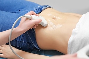
Breast Ultrasound vs Mammogram vs MRI
Three tests, three different jobs. What each one sees, where each falls short, and which is likely to answer your particular question.

These are not three grades of the same test, with MRI at the top. They answer different questions, and the right one depends on your age, your symptom and what has already been done. Choosing well matters more than choosing the most powerful machine.
One thing to be clear about upfront: we offer breast ultrasound only. Mammography and MRI are available through the NHS and hospital breast clinics, and if that is what your situation calls for, we will tell you so rather than scan you anyway.
How the three compare
| Ultrasound | Mammogram | MRI | |
|---|---|---|---|
| How it works | Sound waves, no radiation | Low-dose X-ray | Magnetic field, usually with contrast injection |
| Best at | Telling cysts from solid lumps; assessing a specific area you can feel | Detecting microcalcifications; population screening | Highest sensitivity; extent of known disease; high-risk screening |
| Dense breast tissue | Works well | Harder to read | Works well |
| Typical age | Any age, first-line under 40 | Generally 40 and over | Selected cases only |
| Radiation | None | Low dose | None |
| In pregnancy | Suitable | Usually avoided | Usually avoided |
| Main limitation | Cannot see microcalcifications; not a screening test | Less useful in dense tissue; uncomfortable for some | More false alarms; costly; contrast needed |
| Available at IUS London | Yes | No | No |
Which one answers your question
You can feel a lump and you are under 40. Ultrasound first. Younger breast tissue is denser, which is exactly where mammograms struggle and ultrasound performs well.
You can feel a lump and you are over 40. Usually both, and typically at a breast clinic: the mammogram surveys the whole breast, the ultrasound interrogates the specific area.
You have no symptoms and you are 50 to 71. The NHS breast screening programme invites you for mammography roughly every three years. That is the test with evidence behind it for screening — ultrasound is not a substitute, and going private instead of attending is a poor trade.
You were told you have dense breasts. Ultrasound can add a second look at tissue a mammogram reads less well. It is an addition, not a replacement.
You have a strong family history. Start with your GP, not a scan. Risk assessment may lead to genetic testing and, for those at high risk, an NHS surveillance programme that can include MRI from a younger age — a pathway no private ultrasound can stand in for.
Where our scan fits
- 1
A specific concernA lump, a tender area, an armpit swelling — ultrasound is designed for interrogating a place you can point to. 30-minute appointment, no referral, no preparation.
- 2
Speed while you waitIf a clinic appointment is weeks away, a scan can tell you within days whether a lump is a simple cyst — without stopping the referral.
- 3
Honest routingIf your question needs a mammogram, MRI or biopsy, we say so. Our report is written for a breast clinic to act on.
Your questions answered
Is MRI simply the best test?
Can ultrasound replace my NHS mammogram?
Why is ultrasound better for younger women?
Do I need contrast for a breast ultrasound?
What are microcalcifications and why do they matter?
Can I just book all three privately?
Where to go from here
Lumps, pain and changes — and which need your GP first.
Read the guide →
TypesScreening vs DiagnosticTwo different jobs, and which one you actually need.
Read the guide →
DetectionUltrasound and Early DetectionWhat the scan contributes, stated without overselling.
Read the guide →
The right test for the question
Breast ultrasound in a 30-minute appointment, findings explained on screen as we scan and the written report usually within two hours — and a straight answer if what you need is a test we do not offer.
Kensington, London W8 4ED
3 mins from Notting Hill Gate
This article is general information about breast imaging, not individual medical advice. Any new breast change should be assessed by your GP, who can refer you to an NHS breast clinic — see NHS guidance on breast screening. We offer breast ultrasound only; mammography, MRI and biopsy are not available at IUS London.
Written and clinically reviewed by the HCPC-registered sonographers who perform this scan at IUS London — a CQC-registered diagnostic ultrasound clinic (Provider ID 1-2775844974). Your own findings are explained to you at the scan and set out in your report.
Author: Yianni Kiromitis, Senior Sonographer, HCPC RA38415 — over 20 years’ experience in NHS and private ultrasound
Medically reviewed: 9 August 2026