6 Week Pregnancy Scan — What You Can See and What to Expect
Real scan images from six weeks, what each shape on the screen actually is, the measurements that matter — and why some things are not visible yet.
At six weeks, an early pregnancy scan is often the first time you see anything at all — and what appears on screen is far less baby-shaped than most people expect. There is no recognisable baby at this stage: what the sonographer is looking for is a small dark sac, a bright ring inside it, and if you are far enough along, a tiny flicker. This guide shows what that actually looks like, explains each part, and sets out honestly what a six-week scan can and cannot tell you.
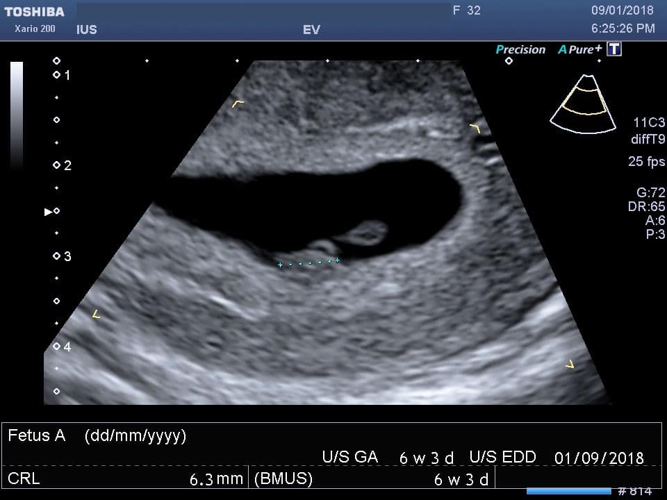
What you can see on a 6 week scan
Three structures appear in sequence during these weeks, and at six weeks you may see all three or only the first one or two — both are normal, because a few days makes a visible difference at this stage.
1. The gestational sac — a black oval. Fluid appears black on ultrasound, so the sac containing the pregnancy shows as a small dark circle or oval within the bright tissue of the womb lining. It is usually the first thing visible, from around five weeks, and its size can be measured to help with dating.

2. The yolk sac — a small bright ring. Inside the gestational sac, a delicate white circle appears from around five and a half weeks. The yolk sac nourishes the embryo before the placenta takes over, and seeing it is an important reassurance: it confirms that what is being looked at is a genuine pregnancy sac rather than fluid.
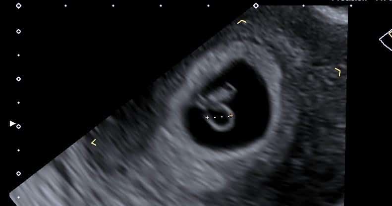
3. The fetal pole and heartbeat. The embryo itself — called the fetal pole at this stage — appears as a small thickening beside the yolk sac, typically from around five and a half to six and a half weeks. The heartbeat, when visible, does not look like a heart: it is a rapid flicker, often described as two fine parallel lines pulsing. It is not always visible at six weeks, and its absence at this stage is frequently just a matter of timing.

What you will not see at six weeks: arms, legs, a face, a profile, or anything resembling the images people associate with baby scans. Those belong to later scans — and a six-week scan that shows a dark sac and a bright ring is doing exactly what it should.
Sizes and measurements at six weeks
| What is measured | Typical at around 6 weeks | Why it matters |
|---|---|---|
| Crown–rump length (CRL) | Roughly 5–9mm | The most accurate way to date a pregnancy at this stage |
| Mean sac diameter (MSD) | Around 15–20mm | Used for dating when no embryo is visible yet |
| Fetal heart rate | Around 100–120 bpm when first seen | Rises quickly over the next fortnight — see rates by week |
A heart rate that seems slow at six weeks is often simply an early rate — rates climb steeply through weeks seven and eight. It is, however, one of the findings that prompts a follow-up scan to confirm the pregnancy is progressing, rather than something to be waved away. Growth over an interval is always more informative than a single measurement.
Reasons for a scan at six weeks
The NHS does not routinely offer a scan this early — the first standard scan is the dating scan at around 12 weeks — unless there is a clinical reason such as pain, bleeding, or a history that warrants earlier assessment, in which case an Early Pregnancy Assessment Unit is the right route. People choose a private early scan for a handful of recognisable reasons:
- Previous miscarriage, and wanting to know sooner this time
- Conception through fertility treatment
- Uncertainty about dates — irregular cycles, or an unknown last period
- Pelvic pain, particularly on one side
- Spotting or light bleeding
- Wanting visual confirmation after a positive test
Behind all of these sits the scan’s most important job, which is often unstated: confirming the pregnancy is in the right place. Seeing a sac within the cavity of the uterus makes an ectopic pregnancy — where the pregnancy implants elsewhere, most often in a fallopian tube — far less likely. That is a genuine clinical answer, and it is the main reason an early scan can be worth having rather than simply reassuring.
Severe or one-sided tummy pain, pain at the tip of your shoulder, heavy bleeding, or feeling faint or dizzy. These can indicate an ectopic pregnancy and need assessment straight away — go to A&E or call 999.
How the scan is done
At six weeks, scanning is normally transvaginal — a slim probe gives a far clearer view of a very small pregnancy than scanning through the abdomen, which at this stage may show almost nothing even when everything is fine. It is not painful, you stay in control throughout, and our first-time guide explains exactly what to expect. No fasting is needed, and unlike later scans you do not need a full bladder for a transvaginal scan — an empty one is more comfortable.
The appointment takes about 30 minutes. Findings are explained to you on screen as they are found, and a written report follows, usually within two hours — written so your GP, midwife or an EPAU can act on it. If you would like to understand the terminology used, our guide to reading an ultrasound report covers the phrases that recur.
When the scan cannot confirm what you hoped
Six weeks sits right at the edge of what ultrasound can resolve, so inconclusive scans are common and are not the same as bad news. If no heartbeat is seen, the commonest explanation is that it is simply too early — our detailed guide to no heartbeat at 6 weeks explains the measurement thresholds that govern the diagnosis and why a repeat scan is arranged rather than a conclusion drawn. If nothing at all is seen in the uterus despite a positive test, that situation has its own name and process, covered in positive test but no baby on ultrasound.
In both cases the next step is the same: a repeat scan after enough time for change to show — usually seven to ten days — because growth over an interval is what answers the question. Booking a second scan two or three days later feels productive but is generally too soon to tell you anything new.
What six weeks really looks like in the room
The second pattern: dates. A meaningful proportion of scans booked at “six weeks” measure a few days behind, because ovulation happened later than the calendar assumed — and those pregnancies go on entirely normally. It is why we measure and, where needed, rescan rather than pronounce on a first look. These are generalised patterns from our practice, not individual cases; your own findings are always explained to you at your scan and set out in your report.
Your questions answered
What does a 6 week ultrasound look like?
Can you see the baby at 6 weeks?
Should there be a heartbeat at 6 weeks?
How big is the baby at 6 weeks on ultrasound?
Is the scan internal or on the tummy?
Do I need a full bladder for a 6 week scan?
Does the NHS do a scan at 6 weeks?
Can a 6 week scan detect twins?
Where to go from here
A week on, and considerably more to see — what changes between six and seven.
Read the guide →
Week by WeekGestational Sac DevelopmentWhat should be visible when — sac, yolk sac, fetal pole, heartbeat.
Read the guide →
If Not SeenNo Heartbeat at 6 WeeksWhy it is usually too early, and the thresholds behind the diagnosis.
Read the guide →
An early pregnancy scan, explained as it happens
Transvaginal scanning by HCPC-registered sonographers, each structure named on screen as it appears, and a written report usually within two hours for your GP, midwife or EPAU.
Kensington, London W8 4ED
3 mins from Notting Hill Gate
Scan images on this page are from examinations performed at IUS London and are shown as illustrative examples of early pregnancy appearances; every pregnancy differs. NHS scanning pathways are described per NHS guidance on pregnancy ultrasound scans. This article is general information, not individual medical advice — urgent symptoms need urgent assessment.
Written and clinically reviewed by the HCPC-registered sonographers who perform this scan at IUS London — a CQC-registered diagnostic ultrasound clinic (Provider ID 1-2775844974). Your own findings are explained to you at the scan and set out in your report.
Author: Yianni Kiromitis, Senior Sonographer, HCPC RA38415
Medically reviewed: 1 August 2026