7 Week Ultrasound — What You See, Day by Day
Week seven is when an early scan starts to reward you. Real images, expected measurements for each day from 7+0 to 7+6, and how to decode the numbers on your own report.
Seven weeks is a noticeably better week to scan than six. The embryo has roughly doubled in length, a heartbeat should now be visible, and the difference between one day and the next is genuinely measurable — which is why so many people search for their exact day rather than just “seven weeks”. This guide covers what appears on screen, what your measurements should be for each day of the week, and what the numbers in your report mean.
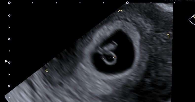
Expected measurements from 7+0 to 7+6
In this window the embryo grows at roughly a millimetre a day, which is what makes day-level precision meaningful. These are typical figures — a few millimetres either side is ordinary variation, and your own report should be read against your dates rather than against a table.
| Gestation | Typical crown–rump length (CRL) | What is usually visible |
|---|---|---|
| 7 weeks + 0 days | About 10mm | Sac, yolk sac, fetal pole, heartbeat expected |
| 7 weeks + 1 day | About 11mm | Heartbeat clearly visible |
| 7 weeks + 2 days | About 12mm | Embryo distinctly separate from the yolk sac |
| 7 weeks + 3 days | About 13mm | Head end becoming distinguishable |
| 7 weeks + 4 days | About 14mm | Early limb buds may be seen |
| 7 weeks + 5 days | About 15mm | Clear curved embryo shape |
| 7 weeks + 6 days | About 16mm | Movement sometimes visible |
The fetal heart rate at seven weeks typically sits somewhere around 120–160 beats per minute, having risen sharply from the slower rates of week six — our guide to fetal heart rate by week sets out the ranges properly.
What your gestational sac measurement means
If your report gives a sac size in millimetres or centimetres and no explanation, there is a long-standing rule of thumb clinicians use as a rough guide: gestational age in days is approximately the mean sac diameter in millimetres plus 30. So a 20mm sac corresponds to roughly 50 days — about 7 weeks and 1 day.
| Mean sac diameter | Approximate gestation |
|---|---|
| 10mm (1.0cm) | About 5 weeks + 5 days |
| 14mm (1.4cm) | About 6 weeks + 2 days |
| 18mm (1.8cm) | About 6 weeks + 6 days |
| 20mm (2.0cm) | About 7 weeks + 1 day |
| 25mm (2.5cm) | About 7 weeks + 6 days |
Two honest caveats. This is an approximation used as a cross-check, not a precise dating tool — and once an embryo is visible, the crown–rump length replaces it entirely, because CRL is considerably more accurate. Sac size matters most when there is no embryo to measure yet. Our gestational sac development guide covers the full week-by-week picture, and the report guide explains the surrounding terminology.
6 weeks versus 7 weeks — the difference a week makes
People often ask whether to scan now or wait, and the honest answer is that a week is a large difference at this stage. At six weeks the embryo measures a few millimetres, the heartbeat may or may not be visible, and an inconclusive result is common. At seven weeks the embryo has roughly doubled, a heartbeat is expected rather than hoped for, and the scan is far more likely to give you a definite answer in one visit.
If you have a choice and no symptoms prompting urgency, waiting until seven weeks usually means fewer repeat scans and less uncertainty. If you have pain or bleeding, do not wait — that is a reason to be assessed now, through an Early Pregnancy Assessment Unit or, for severe symptoms, A&E.
Severe or one-sided tummy pain, pain at the tip of your shoulder, heavy bleeding, or feeling faint or dizzy — go to A&E or call 999, as these can indicate an ectopic pregnancy.
Internal or abdominal — and what about 3D?
Transvaginal scanning remains standard at seven weeks. An abdominal scan may show the pregnancy sac by now, but detail is much poorer — and detail is the entire point of scanning this early. The internal probe enters the vagina only; it does not enter the womb and does not touch the pregnancy, and it is not known to cause any harm in pregnancy. Our first-time guide explains what to expect, and no full bladder or fasting is needed.
3D scanning at seven weeks is not something we offer, and not something worth seeking. At this stage the embryo is a curved shape a centimetre long with no recognisable features, so 3D rendering adds nothing you cannot see in 2D — and we take the position that every scan should earn its place clinically rather than commercially. The scans where surface detail becomes meaningful are much later in pregnancy.
Measuring behind, or no heartbeat yet
Measuring a week behind at seven weeks is a common and usually benign finding — it most often means ovulation happened later than your dates assumed, and your due date is simply revised to match the measurement. Where the embryo measures behind and a heartbeat is present, the pregnancy is generally reassessed at a repeat scan and frequently continues entirely normally. Measurements, not calendars, decide dating.
No heartbeat at seven weeks carries more weight than at six, because by now one is expected. It is not automatically conclusive: the diagnostic threshold under UK guidance is an embryo measuring 7mm or more with no cardiac activity, and even then practice is to confirm with a second opinion or repeat scan. Our guides to no heartbeat at 7 weeks and at 6 weeks set out how that assessment works.
Your questions answered
What does a 7 week ultrasound look like?
What should the CRL be at 7 weeks?
How many weeks is a 20mm gestational sac?
Should there definitely be a heartbeat at 7 weeks?
Is a 7 week scan internal or on the tummy?
Can you see twins at 7 weeks?
Can you do a 3D scan at 7 weeks?
My baby is measuring a week behind at 7 weeks — is that bad?
The week-by-week guides
What is visible a week earlier — and why results are more often inconclusive.
Read the guide →
8 Weeks8 Week Pregnancy ScanLimb buds, movement, and a clearly formed embryo.
Read the guide →
Week by WeekGestational Sac DevelopmentSac, yolk sac, fetal pole and heartbeat — what appears when.
Read the guide →
A seven-week scan, explained as it happens
Transvaginal scanning by HCPC-registered sonographers, measurements taken and explained on screen, and a written report usually within two hours for your GP or midwife.
Kensington, London W8 4ED
3 mins from Notting Hill Gate
Measurements given are typical reference values used as guides; individual variation is normal and your own dating should be interpreted by the clinician reporting your scan. Diagnostic thresholds follow NICE guideline NG126. Scan images are from examinations performed at IUS London and shown as illustrative examples. This article is general information, not individual medical advice — urgent symptoms need urgent assessment.
Written and clinically reviewed by the HCPC-registered sonographers who perform this scan at IUS London — a CQC-registered diagnostic ultrasound clinic (Provider ID 1-2775844974). Your own findings are explained to you at the scan and set out in your report.
Author: Yianni Kiromitis, Senior Sonographer, HCPC RA38415
Medically reviewed: 1 August 2026