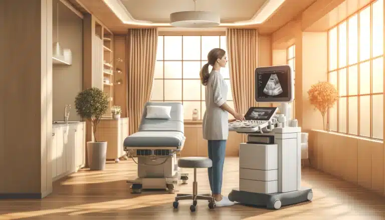
The private abdominal scan is one of our most popular examinations. Upper abdominal pains that can be caused by calculi within the gallbladder are very common.
Cholelithiasis is the medical term for Gallstone disease. Cholelithiasis is one of the most common and costly of all digestive system diseases.
This post outlines some associated risk factors and the more common causes of gallstone formation with some additional details about their classification.
According to the NHS, gallstones are thought to be caused by an imbalance in the chemical makeup of the bile within the gallbladder. These chemical imbalances cause tiny crystals to form within the bile that can gradually increase in size from tiny grains of sand to the size of a pebble over a period of time.
Risk Factors
The risk factors identified by Wang and Afdhal (2016) for gallstones in the gallbladder (cholelithiasis) include:-
- diet,
- age,
- gender,
- oestrogen therapy,
- obesity,
- fasting,
- diabetes,
- family history,
- rapid weight loss,
- some medications including those that reduce cholesterol or Lipids or
- an antibiotic called Cerfriaxone,
- disease of the ilium or it’s resection and
- spinal cord injuries.
Stockley (2001) states that gallstones are not exclusive to fair, fat, flatulent, fertile over 40 years old females as was previously thought but are also found in young and old alike and have even been detected on fetal ultrasound scanning in the womb.
According to Nathanson (2014) it has been estimated from autopsy studies that 12% of men and 24% of women of all ages have gallstone disease present and that 10-30% of them become symptomatic. There are over 40,000 operations to remove the gallbladder and its gallstones (cholecystectomy) performed annually in the UK.
Stockley (2001) states that gallstones are formed in several ways:
- Cholesterol stones which are hard are formed due to an increase in the concentration of cholesterol in the blood (hypercholesterolaemia).
- An increase in bilirubin in the blood (hyperbilirubinaemia) found in patients with haemolytic anaemia which form irregularly shaped soft, small brown pigment gallstones.
- Biliary stasis caused by a faulty, malformed, non-emptying gallbladder or obstructed cystic duct leading to stagnant bile. This creates high concentrations of cholesterol and bile pigments following excessive water absorption. This leads to the formation of mixed cholesterol and bile pigment stones, the most common type of gallstone.
Gallstone Classification
There are different methods used for gallstone classification, namely their
- chemical composition
- location
Wang and Afdhal (2016) classify gallstones into 3 types based on their chemical composition and macroscopic appearance: cholesterol, pigment and rare stones.
- 75% of gallstones in the Western world are cholesterol stones consisting mainly of cholesterol monohydrate crystals and precipitates of amorphous calcium bilirubinate. These stones are further sub-classified as either pure cholesterol or mixed stones that contain at least 50% of cholesterol by weight.
- The remainder of gallstones are classified as pigmented stones that contain mostly calcium hydrogen bilirubinate and they can be further sub-classified into two groups: black pigment (20%) and brown pigment stones (4.5%).
- Rare gallstones account for 0.5% and include calcium carbonate stones and fatty acid-calcium stones.
Wang and Afdhal (2016) classify gallstones by their location as
- Intrahepatic stones which are predominantly brown pigment stones
- Gallbladder stones which are mainly cholesterol stones with a small group of black pigment stones.
- Bile duct stones (choledocholithiasis) which are composed mostly of mixed cholesterol stones.
Gallstone Diagnosis
The abdominal ultrasound scan is the first line of investigation in the diagnosis of gallstones. This ultrasound scan is performed on a fasted patient.
The reason for fasting is that the gallbladder is like a balloon. When we eat something fatty, the gallbladder will excrete the bile into the gut to break down the fat and therefore the gallbladder collapses and it is not possible therefore to see if there are any stones within the lumen.
International Ultrasound Services offers private ultrasound scans to evaluate your gallbladder for any signs of gallstones, thickening of the gallbladder wall and the existence of any pericholecystic fluid. We will also check your liver, your pancreas, your kidneys and the spleen at the same time.
You can book an ultrasound scan in London by visiting our ultrasound scan appointments booking page. You can find more information about the upper abdominal scan here.
References:
NHS Choices (2016) Gallstones causes. Available at: http://www.nhs.uk/Conditions/gallstones/Pages/causes.aspx [Accessed 17/10/2016]
Wang, D., Afdhal, N. (2016). Gallstone Disease In: Feldman, M., Friedman, L., Brandt,L.(eds) Sleisanger and Fordtran’s Gastrointestinal and Liver Disease Pathophysiology / Diagnosis / Management. Volume 1. 10th Edition. Philadelphia, Saunders Elsevier. pp -1100 - 1108
Stockley, M (2001) Abdominal Ultrasound. 1st edition. Greenwich Medical Media
Nathanson, L. (2014) Gallstones, In: Garden, O., Parks, R. (eds.) Hepatobilary and Pancreatic Surgery. A Companion to Specialist Surgical Practice. 5th edition. Edinburgh. Saunders Elsevier. p 174.
Bibiliograph:
http://www.webmd.com/digestive-disorders/gallstones#1What Are Gallstones? [Accessed 17/10/2016]
http://www.livescience.com/34726-gallstones-symptoms-treatment.html. [Accessed 18/10/2016]
Mayo Clinic (2013) Gallstones causes. Available at: http://www.mayoclinic.org/diseases-conditions/gallstones/basics/causes/con-20020461. [Accessed 18/10/2016]
Medically Reviewed by Tareq Ismail Pg(Dip), BSc (Hons)


