Gallstones on Ultrasound — Appearance, Symptoms and When to Scan
Ultrasound is the method of choice for finding gallstones. Here is what they actually look like on screen, what your report’s terms mean, whether stones can be missed — and the symptoms that should prompt a scan.
Gallstones are hardened deposits of digestive fluid that form in the gallbladder — the small, pear-shaped organ under the liver that stores bile and squeezes it out to digest fat. Many cause no symptoms at all; others announce themselves with the unmistakable pain of biliary colic after a fatty meal. Either way, when gallstones are the question, abdominal ultrasound is the answer’s first test: no radiation, around 30 minutes, and the standard diagnostic used across the NHS and private care alike — the examination itself is our abdominal ultrasound scan.
What do gallstones look like on ultrasound?
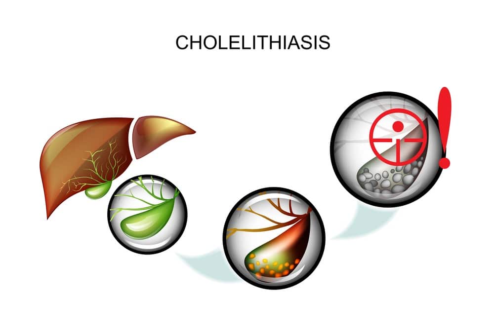
On screen, gallstones are distinctive: bright (echogenic) curved structures inside the gallbladder that cast a dark “acoustic shadow” behind them — the stone reflects almost all the sound, so nothing returns from beyond it. Stones typically move when you change position on the couch, which is one of the checks your sonographer performs. The radiological term for gallstones is cholelithiasis, and that is the word you will usually see in a report.
Ultrasound sees more than the stones themselves. Biliary sludge appears as low-level echoes layering in the gallbladder without a shadow — thickened bile that can cause identical symptoms and precede stones. An inflamed gallbladder (cholecystitis) shows a thickened wall, sometimes fluid around it, and focal tenderness exactly where the probe presses — the sonographic Murphy’s sign. And the scan measures the bile ducts: a dilated duct raises the question of a stone that has escaped the gallbladder and is blocking the drainage system.
Reading a gallbladder ultrasound report
The phrases that appear most often, translated: “cholelithiasis” — gallstones are present (the report will usually note the largest stone’s size and whether there are single or multiple stones). “Biliary sludge” — thickened bile, managed on symptoms. “Gallbladder wall thickening” — possible inflammation, read together with your symptoms and the Murphy’s sign. “CBD not dilated” — the common bile duct measures normal, reassuring against an escaped stone. “Gallbladder contracted” — often simply means you weren’t fasted, which is why preparation matters.
Our reports translate these findings in plain English at the scan itself, and the wider vocabulary is covered in our guide to understanding your abdominal ultrasound report.
Can gallstones be missed on ultrasound?
Occasionally, yes — and an honest page says so. Ultrasound is highly accurate for stones inside the gallbladder, but very small stones and sludge can sometimes escape notice, and stones that have moved into the bile ducts are genuinely harder to see because overlying bowel gas obscures the lower duct. A gallbladder that is contracted (not fasted) also hides its contents — one more reason the four-hour fast matters.
If your scan is normal but typical biliary pain continues, that result is still useful evidence — and the next step belongs with your GP: a repeat fasted scan, blood tests, or specialised imaging of the ducts (MRCP or endoscopic ultrasound, arranged through a specialist) all build on the report you already hold.
Gallstone symptoms — when a scan is worth booking
The hallmark is biliary colic: a steady, intense pain in the upper right abdomen or centre-top of the tummy, classically an hour or so after a fatty meal, often radiating to the back or right shoulder blade, lasting from thirty minutes to several hours, sometimes with nausea. Between attacks you may feel completely well. Less specific patterns — recurring indigestion, bloating after eating, intolerance of fatty food — overlap with other causes, which is exactly what the scan untangles; our guide to stomach pain after eating maps the possibilities.
Pain with fever or chills, yellowing of the skin or eyes (jaundice), dark urine with pale stools, persistent vomiting, or severe pain that will not settle can signal an inflamed gallbladder, a blocked duct or pancreatitis — go to A&E or call 999. A booked scan is never a reason to delay emergency care.
The pattern we see most
The reverse matters too: plenty of scans confirm a healthy, stone-free gallbladder — and that documented exclusion redirects attention to the real culprit rather than leaving “probably gallstones” hanging unproven. These are generalised patterns from our imaging casebook, not individual cases; your own findings are always discussed with you at your scan.
Gallstones and ultrasound — your questions answered
Do gallstones show up on ultrasound?
How should I prepare for a gallbladder ultrasound?
Does the size of a gallstone matter?
Can gallstones be missed on an ultrasound?
Do gallstones always need surgery?
How is this diagnosed on the NHS, and why scan privately?
Where to go from here
The gallstone examination — liver, gallbladder, pancreas, spleen, kidneys and aorta in one visit.
About the scan →
SymptomStomach Pain After EatingGallbladder, stomach or something else — the post-meal pain map.
Read the guide →
Your ReportAbdominal Report, ExplainedThe full vocabulary of an abdominal ultrasound report, translated.
Read the guide →
Is it your gallbladder? Find out this week.
A 30-minute fasted abdominal ultrasound, findings shown to you on screen, and a written report — usually within two hours — that your GP or surgeon can act on.
Kensington, London W8 4ED
3 mins from Notting Hill Gate
Clinical context on gallstones reflects NHS guidance on gallstones. This article is general information, not individual medical advice — management decisions belong with your GP or surgeon, and our reports are written to support exactly those decisions.
Written and clinically reviewed by the HCPC-registered sonographers who perform this scan at IUS London — a CQC-registered diagnostic ultrasound clinic (Provider ID 1-2775844974). Your own findings are explained to you at the scan and set out in your report.
Author: Yianni Kiromitis, Senior Sonographer, HCPC RA38415
Medically reviewed: 31 July 2026