Ultrasound and Cancer Screening — What It Can and Can’t Detect
Ultrasound finds and characterises lumps extremely well. It is not a whole-body cancer check — and no honest clinic will sell it as one. Here is the real map.

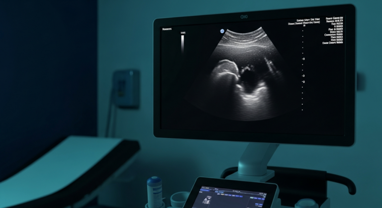
Two things are true at once. Ultrasound is often the first and best test for a lump you can feel — it shows in real time whether a lump is solid or fluid, how it is shaped, and whether blood flows through it, which is exactly the information that separates “benign, be reassured” from “this needs a biopsy”. And at the same time, ultrasound is not a cancer-screening test: it cannot survey the whole body, it cannot see into gas-filled organs like the stomach and bowel or air-filled lungs, and no scan of any kind can tell you that you do not have cancer anywhere. Understanding both halves is what makes a scan genuinely useful — and this page covers the NHS screening programmes you should always attend, what ultrasound contributes, and the questions we are asked about it every week.
Cancer screening belongs to the NHS programmes
Cancer screening means testing people without symptoms to catch specific cancers early, and the UK runs three national programmes: cervical screening (smear tests, broadly ages 25–64), breast screening (mammograms, broadly ages 50–70), and bowel screening (home stool-sample kits, with invitations from your 50s). None of these uses ultrasound as its screening test, all are free, and every invitation is worth taking up — current age ranges and details are on the NHS screening pages.
One rule outranks every screening schedule: screening is for people with no symptoms. If you have a symptom — a lump, bleeding, unexplained weight loss — do not wait for an invitation. See your GP promptly, and use imaging to answer the specific question your symptom raises. That is where ultrasound earns its place.
What ultrasound genuinely contributes
Characterising a lump you can feel. Solid or cystic, smooth or irregular, avascular or blood-fed — these features, visible live on screen, decide whether a lump can be confidently called benign or needs biopsy. This is ultrasound’s core cancer-related job, and it does it anywhere a probe can reach: neck, armpit, breast, groin, skin and soft tissue, testes.
Organ-specific strengths. For testicular lumps ultrasound is the definitive first test. For thyroid nodules it risk-stratifies which few need a needle. For breast lumps it complements mammography — particularly in younger women and dense breast tissue. In the pelvis it assesses ovarian findings, often alongside a CA-125 blood test. And in the abdomen it finds and measures liver and kidney masses, though characterising them fully sometimes needs CT or MRI.
Guiding what happens next. Because the image is live, ultrasound guides biopsies precisely — and because our reports state findings with honest urgency, a suspicious result moves fast: routed to your GP for an urgent referral, with the imaging evidence the next clinic needs already written down.
What ultrasound cannot do
It cannot see through gas or air — so the stomach lining, most of the bowel, and the lungs are out of reach; those organs belong to gastroscopy, colonoscopy and CT. It cannot detect microscopic disease or say a lump is definitely cancer — a suspicious finding still needs tissue diagnosis by biopsy. It cannot survey the whole body in one pass; every examination answers a targeted question about a specific region. And a normal scan of one area is exactly that — reassurance about that area, not a clean bill of health.
That is also the honest answer to “is a private scan better at detecting cancer than the NHS?” — the physics is identical. What differs is access: how quickly you are scanned, how long the appointment lasts, and how fast the report reaches the person who acts on it. Speed and attention are real advantages; detection magic is not on the menu, here or anywhere.
Ultrasound and cancer — your questions answered
Can an ultrasound tell if a lump is cancerous?
What is the difference between a cyst and a tumour on ultrasound?
Does cancer show up on an ultrasound?
Does colour (red) on the ultrasound screen mean cancer?
What is the best scan to detect cancer?
Can ultrasound detect stomach or bowel cancer?
Can ultrasound detect testicular cancer?
Can you see lymphoma or cancer in lymph nodes on ultrasound?
What honest lump assessment looks like
That routing discipline — explained in our follow-up care standard — is the point of scanning a lump early: benign answers end anxiety quickly, and the rare serious one gains weeks. What we will not do is sell scanning as a cancer guarantee, because no honest clinic can.
Where to go from here
A lump you can feel, characterised on screen — anywhere on the body.
About the scan →
ChoosingUltrasound vs MRI vs CTWhich imaging answers which question — the honest comparison.
Read the guide →
SymptomArmpit Lump — What Is It?Lymph node, lipoma or cyst — how the scan tells them apart.
Read the guide →
A lump deserves an answer, not weeks of wondering
Characterised on screen at your appointment, explained in proportion, and reported in writing — usually within two hours — for your GP to act on.
Kensington, London W8 4ED
3 mins from Notting Hill Gate
NHS screening programme details are drawn from NHS screening (age ranges summarised broadly; check the NHS pages for current criteria). This article is general information, not individual medical advice — symptoms should always go to your GP promptly, and NHS screening invitations should always be taken up.
Written and clinically reviewed by the HCPC-registered sonographers who perform this scan at IUS London — a CQC-registered diagnostic ultrasound clinic (Provider ID 1-2775844974). Your own findings are explained to you at the scan and set out in your report.
Author: Yianni Kiromitis, Senior Sonographer, HCPC RA38415
Medically reviewed: 31 July 2026