No Heartbeat at 6 Weeks — What It Means and What Happens Next
At six weeks, not seeing a heartbeat often means it is simply too early. Here is what that finding does and does not tell you, how the diagnosis is properly made, and what happens now.
If you have just had a scan at around six weeks and no heartbeat was seen, you are almost certainly reading this in a difficult, uncertain moment. So the most useful thing we can tell you first is this: at six weeks, the commonest explanation by far is that it is simply too early to see one — and being asked to come back in a week or two is not bad news being softened. It is the correct, deliberate way this is assessed everywhere in the UK, precisely so that no viable pregnancy is ever wrongly written off.
That uncertainty is genuinely hard to sit with, and we are not going to pretend otherwise. What follows is the honest picture: why a heartbeat may not be visible yet, the criteria doctors and sonographers actually use before anything is called non-viable, what the waiting period is for, and where to get both care and support.
Severe or one-sided tummy pain, pain at the tip of your shoulder, heavy vaginal bleeding, feeling faint, dizzy or collapsing, or pain when opening your bowels or passing urine alongside bleeding. These can be signs of an ectopic pregnancy, which needs assessment straight away — go to A&E or call 999. Do not wait for a booked scan.
Why a heartbeat may not be seen at six weeks
Your dates may be different from your development — this is the commonest reason. Pregnancy is dated from the first day of your last period, which assumes ovulation happened around day 14. In practice, ovulation varies widely between women and between cycles: implantation timing, an irregular or longer cycle, or simply a later ovulation can leave you a week or more “behind” your calculated dates. A pregnancy measuring five weeks when the calendar says six and a half is developing perfectly normally — it is the calendar that is out, not the pregnancy. This is why what the scan measures matters far more than what the dates say.
The type of scan makes a real difference. A transvaginal scan places the probe much closer to the uterus and sees detail one to two weeks earlier than scanning through the abdomen. At six weeks, an abdominal scan may show very little even in a completely healthy pregnancy, which is why transvaginal scanning is standard this early — our first-time guide explains what it involves.
Normal biological variation. Cardiac activity typically becomes visible somewhere between about five and a half and six and a half weeks, and embryos do not all reach the same milestone on the same day. A few days either side of the average is ordinary human variation, not a warning sign.
Practical and technical factors. The position of the uterus (a tilted or retroverted uterus sits further from the probe), fibroids, previous abdominal surgery, or scanning conditions can all make a very small embryo harder to see clearly. These are limitations of the view obtained, not findings about the pregnancy — and an honest report says which it is.
And, honestly: sometimes a heartbeat is not seen because the pregnancy has stopped developing — an early miscarriage. That possibility is real, and this page does not pretend otherwise. But at six weeks it cannot be distinguished from the explanations above on a single scan, which is exactly why the criteria below exist.
How the diagnosis is actually made — and why you are asked to wait
This is the part that is often left unexplained, and it is the part that helps most. UK practice follows national guidance (NICE) that sets deliberately cautious measurement thresholds before any pregnancy is described as non-viable. Two measurements do the work:
- 1
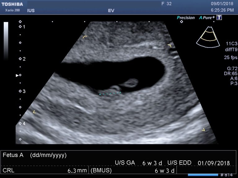
Crown–rump length (CRL) — the embryo’s measurementIf an embryo measures 7mm or more and no heartbeat is seen, that meets the threshold for a diagnosis — and even then, UK practice is to confirm it, usually with a second opinion or a repeat scan. If the embryo measures less than 7mm with no heartbeat, the scan is inconclusive, not negative: a repeat scan is arranged, normally at least seven days later.
- 2
Mean sac diameter (MSD) — the pregnancy sacIf the sac measures 25mm or more with no visible embryo inside, that meets the threshold for an anembryonic pregnancy (sometimes called a blighted ovum). Below 25mm with no embryo yet, the scan is again inconclusive and a repeat is arranged. Our guide to gestational sac development week by week covers what should be visible when.
At six weeks, most pregnancies are well below both thresholds — which is why a six-week scan showing a sac but no heartbeat very often cannot conclude anything at all. The waiting period is not delay or evasion; it is the safeguard. These thresholds were set high on purpose, after evidence showed that tighter criteria risked occasionally misdiagnosing pregnancies that were in fact developing normally. The wait exists so that no one is ever told the wrong thing.
What the repeat scan looks for is change: a sac that has grown, an embryo that has appeared, a heartbeat that is now visible. Growth over time is far more informative than any single snapshot — and where the picture is still unclear, blood tests measuring hCG levels over 48 hours may be used alongside the scan.
When the heart develops and when it becomes visible
The embryonic heart begins as a simple tube and starts beating remarkably early — around three weeks after conception, well before it can be seen on any scan. Visibility, not existence, is what a six-week scan is testing.
| Gestation | What is typically visible on a transvaginal scan |
|---|---|
| 4–5 weeks | A gestational sac may be seen; often nothing more |
| 5–5.5 weeks | Gestational sac with a yolk sac inside |
| 5.5–6.5 weeks | A fetal pole appears; cardiac activity usually becomes visible in this window |
| 7–8 weeks | A heartbeat is expected to be clearly visible; the embryo is measurably larger |
Heart rate also changes rapidly across these weeks — roughly 100–120 beats per minute when first detectable at five to six weeks, rising to around 140–170 by eight to nine weeks. A rate that seems “slow” at six weeks is often simply an early rate, and our guide to normal fetal heart rate by week sets out the ranges. If you are further along than six weeks, the sibling guides for seven weeks and eight weeks address those windows specifically, where the picture is clearer and the thresholds bite sooner.
Where to go and what to expect
The NHS Early Pregnancy Assessment Unit (EPAU) is the right place for this. EPAUs specialise in exactly this situation — scanning, hCG blood tests, and continuity of care through whatever the outcome turns out to be. Many accept self-referral; otherwise your GP, midwife or NHS 111 can refer you. If you are bleeding heavily or in severe pain, go to A&E instead.
A private scan can have a place alongside that pathway — usually when the NHS repeat interval is longer than you can bear to wait, or you want a second look between appointments. It is worth being clear-eyed about what it can offer: an earlier look, not an earlier answer. If the measurements are still below the thresholds above, a private scan will reach the same honest conclusion — that it is too early to say — and repeated scanning a few days apart cannot change what is developing. What it can do is confirm growth when there has been enough time for growth to show.
Whichever route you take, ask for the measurements to be written down: the sac size, the CRL if there is one, and the date. Those numbers are what makes the next scan meaningful, because the comparison is the diagnosis.
Honest words about the outcome nobody wants
Sometimes the repeat scan confirms that the pregnancy has ended. If that happens, a few things are worth knowing plainly. Early miscarriage is common — the NHS estimates around one in eight pregnancies known to the woman end this way, and the true figure including very early losses is higher. It is almost never anyone’s fault: the large majority are caused by chromosomal changes in the embryo that were present from the start, and nothing you did, ate, lifted or felt caused it. Having one early miscarriage does not mean you will have another.
You will be offered choices about what happens next — waiting for things to happen naturally, medication, or a short procedure — and there is usually no rush to decide. Our guide to early ultrasound and miscarriage covers the process in more detail.
And support exists specifically for this. Tommy’s runs a dedicated pregnancy loss support line staffed by midwives, and we keep a list of baby loss helplines including organisations offering counselling and peer support. Using them early is not an overreaction — this is a bereavement, and it deserves to be treated as one.
What we see week to week
We also see the other outcome, and we tell people plainly when the measurements meet the criteria, rather than leaving them to interpret a hedge. What we will not do is scan someone repeatedly over a few days when the interval is too short to show change — that is charging for uncertainty. These are generalised patterns from our practice, not individual cases; your own findings are always explained to you at your scan and set out in your report.
Your questions answered
Is no heartbeat at 6 weeks always bad news?
How long do I have to wait for a repeat scan, and why so long?
What measurements mean a pregnancy is not viable?
Can my dates be wrong even if I know when my last period was?
Would a transvaginal scan see more than the scan I had?
Should I book a private scan while I wait?
Where should I be seen for this on the NHS?
Did I cause this?
If your situation is slightly different
The same question a week on, where the thresholds start to give clearer answers.
Read the guide →
No Sac SeenPositive Test, Nothing on ScanWhen the scan shows no pregnancy in the uterus at all — what that means.
Read the guide →
Week by WeekGestational Sac DevelopmentWhat should be visible when — sac, yolk sac, fetal pole, heartbeat.
Read the guide →
If a repeat scan would help
We scan early pregnancy transvaginally, take the measurements that matter, and tell you plainly what they do and do not show — including when the honest answer is that it is still too early. NHS EPAU care remains the right route for ongoing assessment, and our report is written for them.
Kensington, London W8 4ED
3 mins from Notting Hill Gate
Diagnostic thresholds described here follow NICE guideline NG126 (ectopic pregnancy and miscarriage: diagnosis and initial management). Miscarriage context and figures are from NHS guidance on miscarriage; ectopic warning signs from NHS guidance on ectopic pregnancy. Pregnancy loss support is available from Tommy’s and the organisations listed on our baby loss helplines page. This article is general information, not individual medical advice — your own care should be guided by the clinicians assessing you, and urgent symptoms need urgent assessment.
Written and clinically reviewed by the HCPC-registered sonographers who perform this scan at IUS London — a CQC-registered diagnostic ultrasound clinic (Provider ID 1-2775844974). Your own findings are explained to you at the scan and set out in your report.
Author: Yianni Kiromitis, Senior Sonographer, HCPC RA38415
Medically reviewed: 1 August 2026