
Male Breast Ultrasound in London
Chest swelling or a lump under the nipple, assessed properly — glandular tissue distinguished from fat, and anything that needs a specialist named plainly. Handled without fuss.
- 30-minute appointment — unhurried, where most clinics allow 20
- Glandular tissue distinguished from fatty tissue
- One side or both sides — the armpit lymph nodes are assessed too
- Findings at the scan; written report usually within two hours
- No GP referral required — self-refer today
CQC-registered clinic · Provider ID 1-2775844974 · HCPC-registered sonographers
What is a male breast ultrasound?
A male breast ultrasound examines the tissue behind and around the nipple, the chest wall beneath it, and the lymph nodes in the armpit. It answers a question that cannot be settled by feel alone: is the swelling glandular tissue, or is it fat?
That distinction has a name on each side. True glandular enlargement is gynaecomastia; fatty enlargement without gland is often called pseudogynaecomastia. They feel similar and look similar in the mirror, but they have different causes and different treatment paths — which is why the scan is worth doing rather than guessing.
The great majority of what we find is benign. Male breast cancer exists but is uncommon, and the scan is also there to identify the features that need a specialist rather than reassurance.
Why might I need a male breast scan?
Most men book because something has changed: a firm disc under one nipple, tenderness, or a chest shape that has not responded to training. Gynaecomastia is common at puberty, common again with age, and can be triggered by medication, alcohol, anabolic steroid use, liver problems or low testosterone.
The scan does two things at once. It tells you what the tissue actually is — which decides whether the answer is time, a medication review, or a surgical opinion — and it checks for the small number of findings that need assessing further.
- A firm disc or lump under one or both nipples
- Chest swelling that has not shifted with training
- Tenderness or soreness around the nipple
- A change on one side only
- Swelling since starting a new medication
- Concern about a lump you have found yourself
- Glandular tissue — present, and how much
- Fatty tissue, distinguished from gland
- Comparison between the two sides, where both are scanned
- Any discrete lump, measured and characterised
- Skin and the tissue directly behind the nipple
- Lymph nodes in the armpit
A hard, fixed lump away from the nipple, skin dimpling or puckering, nipple retraction, or bloody discharge should be assessed by your GP promptly — who can refer you to a breast clinic where examination, imaging and biopsy happen together. Male breast cancer is uncommon, but those features are the ones that warrant the specialist pathway rather than reassurance.
Is this scan suitable for me?
A male breast ultrasound is a good fit if any of these sound like you:
- ✓You have noticed a firm disc or lump under the nipple
- ✓Chest swelling has not responded to training or weight loss
- ✓Only one side has changed
- ✓Swelling started after a new medication or supplement
- ✓You want to know whether it is gland or fat before considering surgery
- ✓You would rather have it looked at than keep wondering
And when it is not the right booking
You want the tissue removed — we are a diagnostic clinic and do not perform surgery. If a surgical opinion is the next step, the report gives a surgeon what they need.
You want hormone testing — blood tests for testosterone, thyroid and liver function go through your GP. We do not offer blood tests.
You have a hard fixed lump with skin or nipple change — see your GP promptly for the breast clinic pathway — that route includes biopsy, which we do not provide.
If a male breast ultrasound is not the right test for you, we will say so before you book — not after.
What happens on the day
- 1
Booking — no referral, no preparationNothing to fast for. The appointment is 30 minutes and you can book directly.
- 2
The awkwardness, handled in the first minutePlenty of men put this off because the appointment feels embarrassing. It is a chest scan. You keep your dignity, a chaperone is available on request, and we get on with it without commentary.
- 3
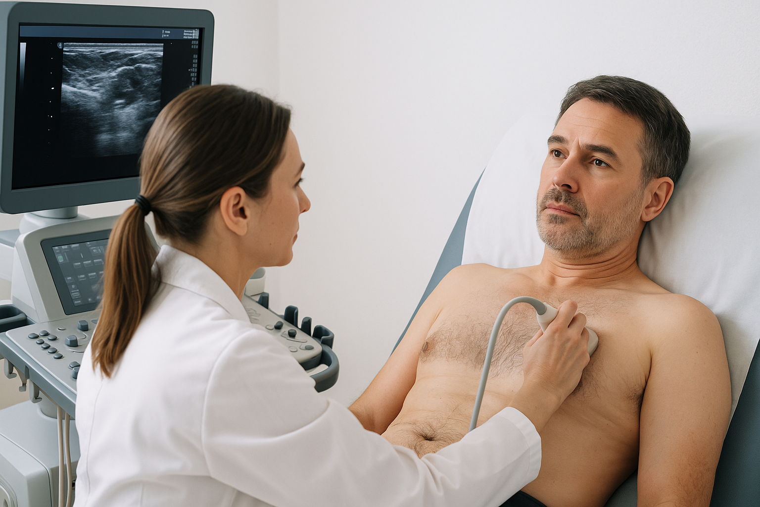
The scanWarm gel and a small probe over the tissue behind and around the nipple, then the armpit. It takes a few minutes per side.
- 4
One side or bothOne side is £189; both sides £269 (£189 + £80), selected when you book. Comparison between sides is what makes a one-sided change interpretable, so call us if you are unsure which to choose.
- 5
Findings explained on screenGland or fat, and what any lump is. You leave knowing the answer rather than waiting for a letter.
- 6
Your reportUsually with you within two hours, written so your GP or a surgeon can act on it directly.
Understanding male chest swelling
Two different tissues, two different answers.
Gynaecomastia — glandular
Actual breast gland tissue behind the nipple, often felt as a firm rubbery disc and frequently tender. Common at puberty and again with age, and sometimes triggered by medication, alcohol, liver problems, low testosterone or anabolic steroid use.
Pseudogynaecomastia — fatty
Fat rather than gland, distributed more diffusely and usually not tender. It tends to respond to weight change where true gland does not, which is exactly why knowing which you have matters before deciding what to do.
The uncommon but important group
Male breast cancer is rare but real. The features that raise concern are a hard fixed lump away from the nipple, skin dimpling, nipple retraction or bloody discharge — and those belong on the breast clinic pathway through your GP.
What your report might say
A normal scan does not override a lump your doctor is concerned about on examination. We do not perform biopsy or mammography, and imaging is one part of an assessment rather than the whole of it.
Gland or fat — know in half an hour
Breast tissue and armpit nodes assessed and explained on screen as we scan, with your written report usually within two hours. One side £189, both sides £269.
How we scan male chests
Scanned by the person reporting it
The sonographer who scans you writes your report. Nothing goes to a remote reader.
No commentary, no fuss
It is a chest scan and we treat it as one. A chaperone is available on request and you can stop at any point.
Both sides available
Comparison between sides is what makes a one-sided change interpretable. Add the second side for £80 when you book.
Named plainly
Gland or fat, stated clearly — because that single answer decides whether the next step is your GP, a surgeon, or nothing at all.
Male Breast ultrasound — your questions answered
Is it gynaecomastia or just fat?
Is male breast cancer common?
Will training or losing weight fix it?
Can medication cause it?
Do I need a GP referral?
Will there be a chaperone?
Do you offer surgery or removal?
Do you do mammograms or biopsies?
Will you check my hormone levels?
How quickly will I get my results?
Other MSK scans we offer
Lumps, pain and swelling assessed with Doppler blood flow.
See the scan →
LumpsLumps & Bumps Scan
Soft tissue lumps anywhere, identified and measured.
See the scan →
Men’s HealthAll Men’s Scans
The full range of men’s health ultrasound we offer.
See scans →
Further reading: men with breast lumps and armpit lumps.
Where can you get a male breast ultrasound in London?
Our clinic is at 5a Lucerne Mews, Kensington, London W8 4ED — a quiet mews three minutes on foot from Notting Hill Gate, serving Kensington, Notting Hill, Holland Park, Bayswater and central London.
Short-notice appointments are often available, including for people facing a long wait elsewhere. Call 0203 051 6506 for today’s availability.
Getting here
By tube: Notting Hill Gate — three minutes’ walk.
By bus: Routes along Notting Hill Gate and Kensington Church Street stop nearby.
By car: On-street parking nearby — full options in directions & parking.
Stop wondering about it
Breast tissue and armpit nodes assessed in a 30-minute appointment — gland or fat named plainly, explained on screen as we scan, with your report usually within two hours.
Kensington, London W8 4ED
3 mins from Notting Hill Gate
Written and clinically reviewed by the HCPC-registered sonographers who perform this scan at IUS London — a CQC-registered diagnostic ultrasound clinic (Provider ID 1-2775844974). Your own findings are explained to you at the scan and set out in your report.
Author: Yianni Kiromitis, Senior Sonographer, HCPC RA38415 — over 20 years’ experience in NHS and private ultrasound · Reviewed 10 August 2026