Understanding Your Armpit Lump: Lymph Node, Lipoma, or Cyst?
Discovering a lump in your armpit can be a source of significant anxiety. The armpit, or axilla, is a complex anatomical region containing a network of lymph nodes, blood vessels, nerves, and fatty tissue. Given this complexity, various conditions can manifest as a palpable lump, ranging from benign and harmless to those requiring urgent medical attention. At IUS London, a premium private ultrasound clinic in Kensington, we understand the concerns that arise with such discoveries and are dedicated to providing clear, compassionate, and clinically excellent guidance.
This comprehensive guide aims to demystify the common causes of armpit lumps, focusing on the key differentiators between enlarged lymph nodes, lipomas, and cysts. We will delve into the clinical nuances, diagnostic approaches, and when it’s crucial to seek immediate medical evaluation.
⚠ Red Flags (A&E: Accident & Emergency) – When to Seek Immediate Medical Attention
While many armpit lumps are benign, certain signs and symptoms warrant immediate medical evaluation at an Accident & Emergency department. Do not delay seeking help if you experience any of the following:
- Rapidly growing lump: A lump that appears suddenly and grows quickly over days or weeks.
- Severe, unexplained pain: Intense pain in the armpit that is not related to injury or muscle strain.
- Associated fever, chills, or night sweats: These can indicate a systemic infection or more serious underlying condition.
- Redness, warmth, and tenderness spreading from the lump: Suggestive of a severe infection (e.g., cellulitis or abscess) that may require urgent drainage or intravenous antibiotics.
- Lump accompanied by unexplained weight loss or persistent fatigue: These are constitutional symptoms that can be associated with malignancy.
- Skin changes over the lump: Such as dimpling, puckering, ulceration, or changes in skin colour, particularly if they resemble an orange peel texture.
- Fixed or immovable lump: A lump that feels firmly attached to the underlying tissue and does not move freely.
- New nipple discharge or changes in breast tissue: Especially if the lump is in the axilla adjacent to the breast, as this could indicate breast pathology.
If you experience any of these red flags, please seek immediate medical attention. Early diagnosis and intervention can be critical for optimal outcomes.
The Anatomy of an Armpit Lump: Common Culprits
To understand what an armpit lump might be, it’s helpful to consider the structures present in the axilla:
1. Lymph Nodes: These small, bean-shaped glands are part of the immune system, filtering lymph fluid and housing immune cells. They are numerous in the armpit, draining the breast, arm, and upper back.
2. Fatty Tissue: The armpit contains a significant amount of adipose tissue.
3. Skin and Subcutaneous Tissue: Hair follicles, sweat glands, and sebaceous glands are abundant in this area.
4. Muscles, Nerves, and Blood Vessels: While less common to form palpable lumps, issues with these structures can sometimes present as such.
Let’s explore the three most common causes of armpit lumps:
1. Enlarged Lymph Nodes (Lymphadenopathy)
Lymph nodes are often the first responders to infection or inflammation in their drainage area. When they enlarge, it’s typically a sign that your immune system is actively fighting something off. This is known as lymphadenopathy.
Causes of Enlarged Lymph Nodes:
- Infections: This is the most frequent cause. Bacterial infections (e.g., cellulitis, cat scratch disease, folliculitis), viral infections (e.g., common cold, mononucleosis, HIV, shingles), and fungal infections can all lead to reactive lymphadenopathy. The nodes become swollen, tender, and sometimes painful as they fill with immune cells.
- Inflammatory Conditions: Autoimmune diseases like rheumatoid arthritis or lupus can cause generalised lymph node enlargement, including in the axilla.
- Allergic Reactions: Severe allergic reactions can sometimes trigger regional lymph node swelling.
- Vaccinations: Recent vaccinations (e.g., COVID-19, MMR) in the arm can cause temporary enlargement of axillary lymph nodes on the vaccinated side.
- Malignancy: This is the most concerning cause. Lymph nodes can enlarge if they are involved in metastatic cancer (e.g., breast cancer, melanoma, lung cancer) or primary cancers of the lymphatic system (lymphoma). Malignant nodes are often firm, non-tender, and fixed to surrounding tissues. They may also be accompanied by constitutional symptoms.
Clinical Presentation: Reactive lymph nodes are typically soft to firm, mobile, and often tender or painful to touch. They usually resolve as the underlying infection or inflammation subsides. Malignant nodes, conversely, tend to be harder, non-tender, and may feel fixed. The size can vary significantly, from a few millimetres to several centimetres.
2. Lipomas
A lipoma is a benign (non-cancerous) tumour composed of fatty tissue. They are among the most common soft tissue tumours and can occur anywhere on the body where fat cells are present, including the armpit.
Causes of Lipomas: The exact cause of lipomas is unknown, but they tend to run in families, suggesting a genetic predisposition. They are more common in middle-aged adults and can sometimes develop after a minor injury, though the link is not fully understood.
Clinical Presentation: Lipomas are typically:
- Soft and doughy to the touch: They feel like a rubbery lump under the skin.
- Mobile: They can be easily moved with finger pressure.
- Painless: Most lipomas are asymptomatic, though they can become painful if they grow large enough to press on nerves or if they are located in an area subject to friction.
- Slow-growing: They usually grow very slowly over months or years and rarely exceed a few centimetres in diameter.
- Located just beneath the skin: They are subcutaneous.
Lipomas are generally harmless and do not require treatment unless they are causing symptoms (pain, cosmetic concern) or if their diagnosis is uncertain.
3. Cysts
Cysts are sacs filled with fluid, semi-solid material, or air. In the armpit, common types include epidermal cysts (also known as sebaceous cysts) and hidradenitis suppurativa-related cysts.
Causes of Cysts:
- Epidermal Cysts: These arise from hair follicles or blocked sebaceous glands. They occur when skin cells, instead of shedding, move deeper into the skin and multiply, forming a sac that fills with keratin (a protein found in skin and hair). They are very common.
- Hidradenitis Suppurativa (HS): This is a chronic inflammatory skin condition affecting areas with apocrine sweat glands, such as the armpits, groin, and buttocks. It causes painful, deep-seated nodules, abscesses, and sinus tracts that can resemble cysts.
Clinical Presentation:
- Epidermal Cysts: Typically smooth, round, and mobile lumps under the skin. They can range in size from small to several centimetres. They are usually non-tender unless they become inflamed or infected, in which case they can become red, swollen, and painful, sometimes discharging a foul-smelling, cheesy material.
- Hidradenitis Suppurativa: Presents as recurrent, painful, inflamed lumps that can rupture and drain pus. Over time, HS can lead to scarring and the formation of interconnected tunnels (sinus tracts) under the skin. It is often associated with significant discomfort and can have a profound impact on quality of life.
Differential Diagnosis: A Comparative Overview
Distinguishing between these conditions is crucial for appropriate management. Here’s a comparative table summarizing their key features:
| Feature | Enlarged Lymph Node | Lipoma | Cyst (Epidermal/HS) |
|---|---|---|---|
| **Feel** | Soft to firm, rubbery | Soft, doughy, rubbery | Smooth, firm, sometimes fluctuant (fluid-filled) |
| **Mobility** | Mobile (reactive), fixed (malignant) | Highly mobile, slips under fingers | Mobile (epidermal), fixed/deep-seated (HS) |
| **Pain** | Often tender/painful (infection/inflammation) | Usually painless | Painless (uninfected), painful (infected/HS) |
| **Growth Rate** | Rapid (infection), slow (chronic inflammation/malignancy) | Very slow, stable | Slow, can fluctuate in size (epidermal), recurrent (HS) |
| **Location** | Deeper, within lymphatic channels | Subcutaneous fat layer | Within skin/subcutaneous layer |
| **Associated Symptoms** | Fever, chills, weight loss (infection/malignancy) | None | Redness, swelling, discharge (infection/HS) |
| **Skin Changes** | None, unless overlying cellulitis | None | Redness, inflammation, punctum (epidermal), scarring, sinus tracts (HS) |
The Role of Ultrasound in Diagnosis
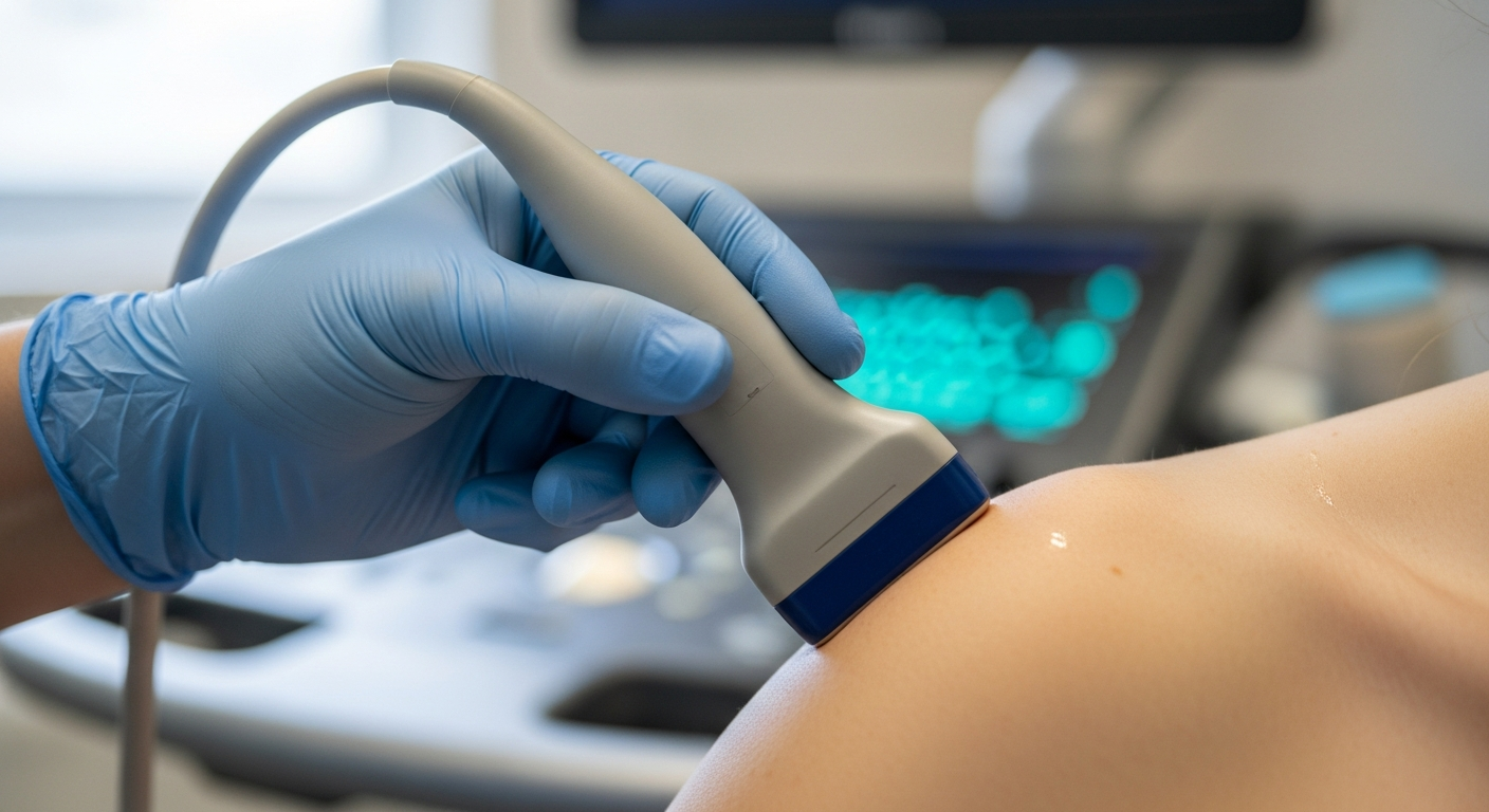
At IUS London, ultrasound imaging plays a pivotal role in the accurate diagnosis and characterisation of armpit lumps. It is a non-invasive, safe, and highly effective tool for visualising soft tissue structures and differentiating between various pathologies.
How Ultrasound Helps:
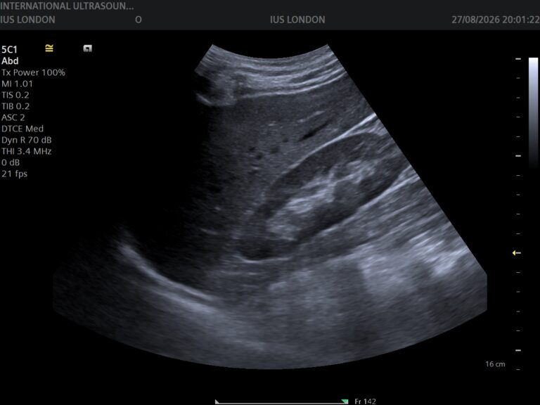
- Detailed Visualisation: Ultrasound provides real-time, high-resolution images of the lump, allowing our expert sonographers to assess its size, shape, borders, internal structure, and relationship to surrounding tissues. This is critical for distinguishing between solid masses (like lymph nodes or lipomas) and fluid-filled structures (like cysts).
- Differentiation of Lymph Nodes: Ultrasound can differentiate between reactive lymph nodes (which typically maintain an oval shape, fatty hilum, and normal blood flow patterns) and suspicious lymph nodes (which may appear rounded, lose their fatty hilum, and show abnormal vascularity, suggesting malignancy) [1, 5]. Advanced techniques like Shear Wave Elastography (SWE) can further assess tissue stiffness, providing additional diagnostic clues for lymph node malignancies [2, 3].
- Characterisation of Lipomas: Lipomas have a characteristic appearance on ultrasound, typically appearing as well-defined, encapsulated, hyperechoic (bright) masses parallel to the skin, often with internal striations. Their soft, compressible nature can also be observed during the scan.
- Identification of Cysts: Cysts are typically seen as well-defined, anechoic (black, fluid-filled) structures with posterior acoustic enhancement. Infected cysts may show internal debris and increased blood flow in the surrounding tissues. For conditions like Hidradenitis Suppurativa, ultrasound can identify the extent of inflammation, abscess formation, and sinus tracts, guiding treatment decisions.
- Guidance for Biopsy: If a lump is suspicious, ultrasound can precisely guide a fine-needle aspiration (FNA) or core needle biopsy, ensuring accurate tissue sampling for pathological examination. This minimally invasive procedure is crucial for definitive diagnosis, especially in cases where malignancy is suspected.
Recent advancements in Artificial Intelligence (AI) integrated with ultrasound have shown promising results in improving the differential diagnosis of superficial soft-tissue masses, including distinguishing between benign and malignant lesions with high accuracy [1]. This technology further enhances the diagnostic capabilities available at clinics like IUS London.
From Our Practice: A Clinical Insight
At IUS London, we frequently encounter patients presenting with armpit lumps. One common clinical pattern we observe involves young to middle-aged women who present with a tender, mobile lump in the axilla, often after shaving or using antiperspirants. Clinically, there’s concern for infection or an inflamed cyst. Ultrasound typically reveals a well-defined, fluid-filled structure consistent with an epidermal cyst or a small abscess, sometimes associated with a blocked hair follicle. In these cases, conservative management with warm compresses and antibiotics is often sufficient, with surgical drainage considered if the abscess is large or persistent. This highlights the importance of detailed clinical history combined with precise ultrasound imaging to avoid unnecessary anxiety and guide appropriate, often conservative, management. [SUGGESTED — verify with clinician]
Conclusion
An armpit lump, while often benign, should never be ignored. Understanding the potential causes—from reactive lymph nodes fighting infection to benign lipomas or inflammatory cysts—is the first step towards peace of mind. The advanced diagnostic capabilities of ultrasound, coupled with expert clinical assessment, are invaluable in accurately characterising these lumps and guiding appropriate management.
At IUS London, our commitment is to provide you with the highest standard of care, utilising state-of-the-art ultrasound technology and the expertise of our medical professionals to ensure a precise and compassionate diagnostic journey.
Take the Next Step for Your Health
If you have discovered an armpit lump and are seeking clarity and expert evaluation, do not hesitate. Early and accurate diagnosis is paramount. Book a private ultrasound scan at IUS London today. Our experienced team is here to provide you with a thorough assessment, answer your questions, and offer personalised care in a comfortable and reassuring environment.
References
[1] Bin Long et al., 2023, Deep learning models of ultrasonography significantly improved the differential diagnosis performance for superficial soft-tissue masses: a retrospective multicenter study, BMC Medicine.
[2] Yujia Gao et al., 2022, Evaluating Different Quantitative Shear Wave Parameters of Ultrasound Elastography in the Diagnosis of Lymph Node Malignancies: A Systematic Review and Meta-Analysis, Cancers.
[3] R. Sigrist et al., 2017, Ultrasound Elastography: Review of Techniques and Clinical Applications, Theranostics.
[4] Xinyang Han et al., 2025, Artificial intelligence performance in ultrasound-based lymph node diagnosis: a systematic review and meta-analysis, BMC Cancer.
[5] D. Fischerová et al., 2021, Terms, definitions and measurements to describe sonographic features of lymph nodes: consensus opinion from the Vulvar International Tumor Analysis (VITA) group, Ultrasound in Obstetrics & Gynecology.
Book Your Private Ultrasound Scan in London
Same-day appointments available in Kensington. No GP referral required. HCPC-registered consultant sonographers.