
Other MSK Ultrasound Scans
When your injury doesn't fit a standard joint page — calf tears, hamstring strains, forearm tendinopathy — our NHS-trained sonographers assess any soft tissue area with the same clinical precision.
- No GP referral required
- Same-day appointments usually available
- Digital report within 2–4 hours
- Fully qualified, experienced NHS sonographers
Your Injury Doesn't Always Fit a Standard Category
Most private ultrasound clinics offer scans for the shoulder, knee, or hip. But what happens when the pain is in your calf after a sprint, your hamstring following a tackle, or your forearm after a repetitive strain injury? These injuries are just as common and just as clinically significant — yet they often fall through the gaps of standard booking systems.
At IUS London, our MSK ultrasound-scan service covers any soft tissue area of the body. Whether you're a weekend runner, a professional athlete, or someone managing a work-related strain, our sonographers assess the specific structure causing your symptoms.
Waiting for an NHS MSK referral takes an average of 4 to 8 weeks after a GP appointment. Untreated soft tissue injuries deteriorate during that window, turning a manageable strain into a complex tear requiring surgical intervention. A private MSK ultrasound-scan at IUS London provides a definitive diagnosis on the same day you book.
- Sudden, sharp pain during activity with an audible pop or snap
- Visible bruising or swelling developing within hours of an injury
- A palpable gap, lump, or indentation in a muscle belly
- Persistent aching or tightness that does not resolve with rest
- Weakness or inability to fully contract a muscle group
- Burning, tingling, or shooting pain suggesting nerve involvement
What Does an MSK Ultrasound-Scan Assess?
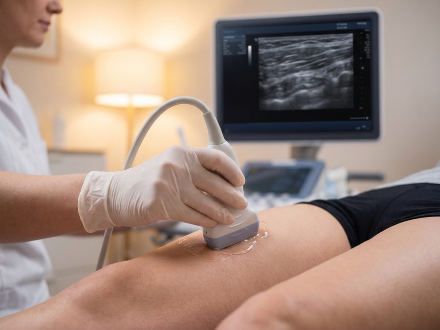
A musculoskeletal ultrasound-scan uses a high-frequency linear transducer to produce real-time, high-resolution images of soft tissue structures. The procedure is non-invasive, uses no radiation, and allows dynamic assessment — the sonographer can observe the tissue as you move, identifying pathology that static imaging would miss.
We assess the gastrocnemius and soleus muscles for partial or complete tears, haematoma formation, and myositis. Calf tears — commonly described as "tennis leg" — produce a characteristic split-screen appearance on ultrasound, showing the fluid-filled gap between the medial gastrocnemius and the soleus aponeurosis.
Hamstring injuries range from mild Grade I strains to complete proximal avulsions at the ischial tuberosity. Ultrasound identifies the precise location and grade of the tear, the presence of a haematoma, and any involvement of the proximal tendon — information that directly determines whether conservative management or surgical repair is appropriate.
The quadriceps muscle group is susceptible to direct contusion injuries and acute strains, particularly at the musculotendinous junction. We assess all four heads — rectus femoris, vastus lateralis, vastus medialis, and vastus intermedius — for tears, calcification, and myositis ossificans.
We assess the distal biceps tendon at the elbow for partial or complete rupture, as well as the triceps tendon for tears at its olecranon insertion. Intramuscular tears and haematomas within the biceps or triceps belly are also clearly visualised.
Repetitive strain injuries affecting the flexor and extensor compartments of the forearm are common in office workers, manual labourers, and racquet sport players. We identify tendinopathy, partial tears, and peritendinous fluid collections, including the common extensor origin at the lateral epicondyle.
Anterior shin pain is a frequent complaint among runners and military personnel. We differentiate tibialis anterior tendinopathy from medial tibial stress syndrome and assess for periosteal oedema and compartment syndrome indicators — conditions that require very different management approaches.
Peroneal tendon pathology is frequently misdiagnosed as a lateral ankle sprain. We assess the peroneus longus and brevis tendons for longitudinal splits, subluxation, and tenosynovitis, as well as the superior peroneal retinaculum for tears that allow the tendons to dislocate over the fibula.
If the area you need assessed is not listed above, contact us before booking. Our sonographers cover virtually any superficial soft tissue structure, including abductor and adductor muscle groups, the iliotibial band, and the popliteal fossa.

How Soft Tissue Injuries Develop and What Ultrasound Reveals
Soft tissue injuries follow predictable mechanical patterns. Understanding which mechanism caused your symptoms helps the sonographer target the correct structures and interpret the findings accurately.
Sudden, forceful eccentric contraction — such as sprinting or jumping — causes muscle fibres to tear at the myotendinous junction. Ultrasound shows a hypoechoic (dark) defect within the muscle belly, often accompanied by a surrounding haematoma. Grading from I (minor fibre disruption) to III (complete rupture) directly determines the rehabilitation timeline.
Repetitive loading without adequate recovery causes micro-tears within the tendon matrix, leading to a degenerative process called tendinopathy. The tendon appears thickened, hypoechoic, and loses its normal fibrillar echotexture. Neovascularisation — new blood vessel ingrowth — is detectable with Doppler and confirms active pathology.
Inflamed bursae appear as anechoic or complex fluid collections on ultrasound. Haematomas following direct trauma show a characteristic evolution from anechoic (acute) to heterogeneous (organising) to hyperechoic (chronic) over days to weeks.
Swollen surrounding tissues compress peripheral nerves, producing burning, tingling, or shooting pain. Ultrasound identifies the site of compression, measures the cross-sectional area of the nerve, and guides targeted treatment decisions.
Your MSK Ultrasound-Scan Appointment, Step by Step
The entire appointment takes approximately 30 minutes. No special preparation is required — wear comfortable clothing that allows access to the area being scanned.
The sonographer discusses your symptoms, the mechanism of injury, and any relevant medical history. This contextual information directs the imaging protocol and ensures the correct structures are assessed.
A clear, water-based ultrasound gel is applied to the skin. The sonographer moves the linear transducer probe over the affected area, systematically evaluating each relevant structure in both longitudinal and transverse planes.
You may be asked to perform specific movements while the sonographer scans. Dynamic assessment reveals pathology that is only apparent under load, such as tendon subluxation or a partial tear that widens with movement.
Where clinically indicated, colour Doppler imaging assesses blood flow within and around the affected tissue. Increased vascularity within a tendon confirms active tendinopathy and guides targeted treatment decisions.
At the end of your appointment, the sonographer provides a clear verbal summary of the findings. You leave the clinic with a definitive understanding of your diagnosis and the recommended next steps.
A comprehensive written digital report is issued within 2 to 4 hours. This report includes the clinical indication, findings, diagnosis, and management recommendations — formatted for sharing with your GP, physiotherapist, or specialist.
From Diagnosis to Recovery: Your Management Pathway
The findings from your MSK ultrasound-scan determine the most appropriate management strategy. A precise diagnosis avoids the common pitfall of generic, unguided rehabilitation that fails to address the actual structural pathology.
-
Conservative ManagementFor Grade I and II strains, the POLICE protocol (Protection, Optimal Loading, Ice, Compression, Elevation) combined with a structured physiotherapy programme produces full recovery in the majority of cases. The scan confirms the grade and guides the loading timeline.
-
Targeted PhysiotherapyYour scan report identifies the specific structure involved — gastrocnemius versus soleus, proximal versus distal hamstring — allowing your physiotherapist to design a rehabilitation programme that targets the correct tissue rather than applying a generic protocol.
-
Orthopaedic or Sports Medicine ReferralGrade III complete ruptures, proximal tendon avulsions, and complex multi-structure injuries require specialist assessment. Your IUS London report provides the referring clinician with the imaging evidence needed to expedite your appointment and plan the appropriate intervention.
-
Return-to-Sport PlanningAthletes and active individuals require objective evidence before returning to full training. A follow-up MSK ultrasound-scan confirms structural healing and guides safe return-to-sport decisions, reducing the risk of re-injury.

When to Book and Common Mistakes to Avoid
Knowing when to seek imaging — and understanding the consequences of waiting — helps you make the right decision for your recovery.
Book Your Scan If You Have:
- Acute muscle pain following a specific injury event
- Chronic tendon pain that has not responded to 4 weeks of rest
- A palpable lump or swelling in a muscle or tendon
- Recurrent injury to the same area despite rehabilitation
- Pre-season assessment before returning to competitive sport
- Your physiotherapist or GP has recommended imaging
What Delays Diagnosis and Worsens Outcomes:
- Waiting for an NHS MSK referral (average 4–8 weeks after GP appointment)
- Continuing to train through a partial tear, converting it to a complete rupture
- Assuming rest alone will resolve a structural tendon injury
- Undergoing generic physiotherapy without a confirmed diagnosis
- Relying on X-ray, which does not image soft tissue structures
A partial muscle tear that is loaded before adequate healing converts to a complete rupture in a predictable pattern. MSK ultrasound-scan provides diagnostic accuracy of 85–95% for soft tissue injuries in experienced hands — accuracy that directly prevents this outcome by confirming the extent of the injury before any rehabilitation loading begins.
Clinical Standards That Match the NHS, Without the Wait
IUS London operates as a dual NHS-private practice. The sonographers who assess your MSK injury are the same clinicians who work in NHS trusts — bringing hospital-grade expertise to a private setting where same-day appointments are the standard, not the exception.
Our clinic is registered and inspected by the Care Quality Commission — the same regulatory body that oversees NHS hospitals. CQC registration confirms our clinical governance meets national standards.
Every sonographer at IUS London holds HCPC registration and has completed formal MSK ultrasound training aligned with the Royal College of Radiologists curriculum. Their dual NHS-private practice means they encounter and correctly interpret complex pathology daily.
Acute injuries require prompt assessment. We offer same-day appointments at our Notting Hill Gate clinic, accessible directly from the Central and Circle lines. No GP referral is required.
Your digital report arrives within 2 to 4 hours of your appointment, formatted for clinical use — including the indication, findings, diagnosis, and management recommendations — so your GP or physiotherapist can act on it immediately.
Our patients consistently highlight the clarity of communication, the professionalism of the sonographers, and the speed of the reporting process. These reviews reflect a service built around patient experience, not just clinical output.
Your MSK ultrasound-scan is £159 with no hidden fees, no referral costs, and no administrative delays. The price you see is the price you pay, and no insurance pre-authorisation is required.
Book Your MSK Ultrasound‑Scan Today
If you are dealing with unexplained muscle pain, a suspected soft tissue injury, or a recurring problem that standard physiotherapy has not resolved, a targeted MSK ultrasound-scan provides the definitive answer. Our team of NHS-trained sonographers at our Notting Hill Gate clinic is ready to assess you — often on the same day you contact us.
Use the booking system to select your preferred date and time. The scan costs £159 and includes your verbal results and a written digital report within 2 to 4 hours.
Book Your Appointment
Select your preferred date and time below. Same-day appointments are available subject to availability.
Dedicated MSK Ultrasound‑Scan Pages
If your concern relates to one of the major joints below, our dedicated scan pages provide condition-specific information and direct booking.