Medical image technology can mean the difference between a complex and uncertain diagnosis and a concrete assurance of a medical condition. Today's imaging clinics are more technologically advanced than ever before, providing patients and doctors with crucial knowledge. It is vital that patients understand the wealth of medical image technology and options that are typically available at any good clinic so that they can be sure they choose the right technique for their needs.
Diagnostic Imaging Modalities
X-Ray Imaging
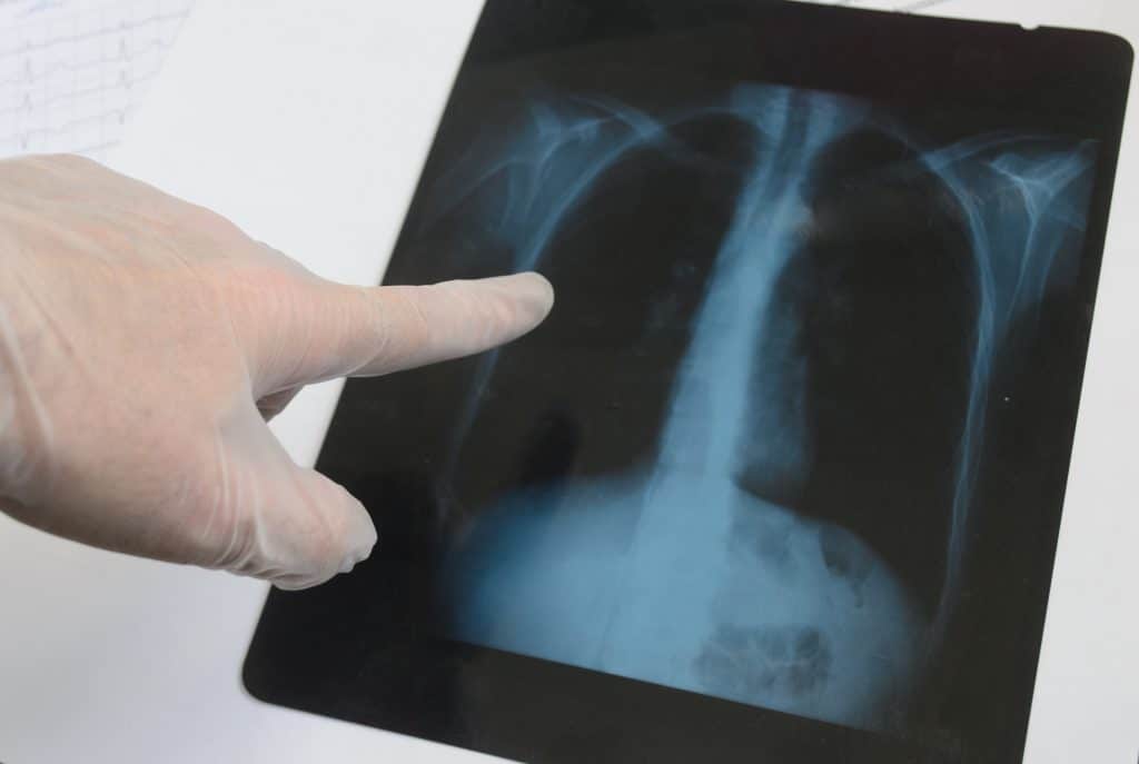
Doctor holding an x-ray
X-ray is the earliest and most common form of diagnostic imaging. In an x-ray, the radiographers use an x-ray generator to take several shots of the body part in question. The x-rays pass through different materials and are being absorbed at different rates, making it possible to take an image of a bone without losing it amidst muscle and other tissues. Bones show up as a strong white, while the rest of the body's tissues appear only faintly. A doctor can use an x-ray image to determine a vast number of problems, ranging from common breaks and fractures to less common ailments.
Ultrasound Imaging
Ultrasounds are well known to most women who have had children. Diagnostic imaging uses ultrasounds to view body structures that cannot be viewed with x-ray technology alone. A developing fetus is not the only type of structure that can be viewed but tendons, muscles, ligaments, complex joint structures, and internal organs can also be imaged with ultrasound.
Ultrasounds measure the reflection of sound to produce images. Typically, this is used to look "beneath" a medium to view the underlying structure. Using the same technology that helps a bat hunt, doctors can use ultrasounds to observe an infant in utero.
You can read more about ultrasound scans.
CT Scans
CT scans, better known to some patients as CAT scans, are another commonly used method of diagnostic imaging. A CT scan uses x-ray technology as well, but combines it with the digital capabilities of a computer. The CT scanner takes multiple frames around a single point from many different angles, and then combines them to reveal the overall structure.
MRIs
Magnetic resonance imaging, or MRI, uses a powerful magnet, radio frequencies, and computer technology to create highly detailed images of organs and other structures within the body. This type of imaging can go so far as to identify infections, joint damage, and diseases of the liver. Other conditions for which you may need an MRI include:
a) Heart problems
b) Brain damage
c) Tumors and cysts
d) Blood flow analysis
e) Diseases of the pancreas
f) Bone & joint pain
h) Reproductive disorders
There are no serious side effects associated with an MRI, but it is important to notify your medical imaging professional about any metal implants, pacemaker, artificial limbs, medical patches, or recently acquired tattoos or other permanent makeup.
Nuclear Medicine
Nuclear medicine is one of the most striking uses of modern image generation technology. In a nuclear medical exam, a slightly radioactive agent is introduced into a patient's system and then monitored to see where it travels. Because the doctor can track the radioactive signal from the compound, he or she can see exactly how whatever the compound was absorbed with is handled by the body. The radioactive isotopes are completely harmless, and will eventually become inert even if it isn't passed from the body shortly after it is introduced.
Imaging Clinics
Almost any imaging clinic in London offers ultrasound scans. Diagnostic imaging centers will sometimes offer CT, x-rays, MRI and mammography,
The usefulness of medical imaging makes it an excellent diagnostic and curative tool for conditions faced by millions of patients each year