Finding a lump in your chest or near the nipple can be unsettling. In my clinical imaging practice, I see many men who assume the worst— but the reality is that most male breast lumps are benign. The key is to assess the pattern of the lump and any associated symptoms, then use symptom-led imaging where appropriate to reach a clear, safe conclusion.
This guide explains the common causes, when you should seek assessment, and how ultrasound supports a symptom-led diagnostic pathway.
Key takeaways
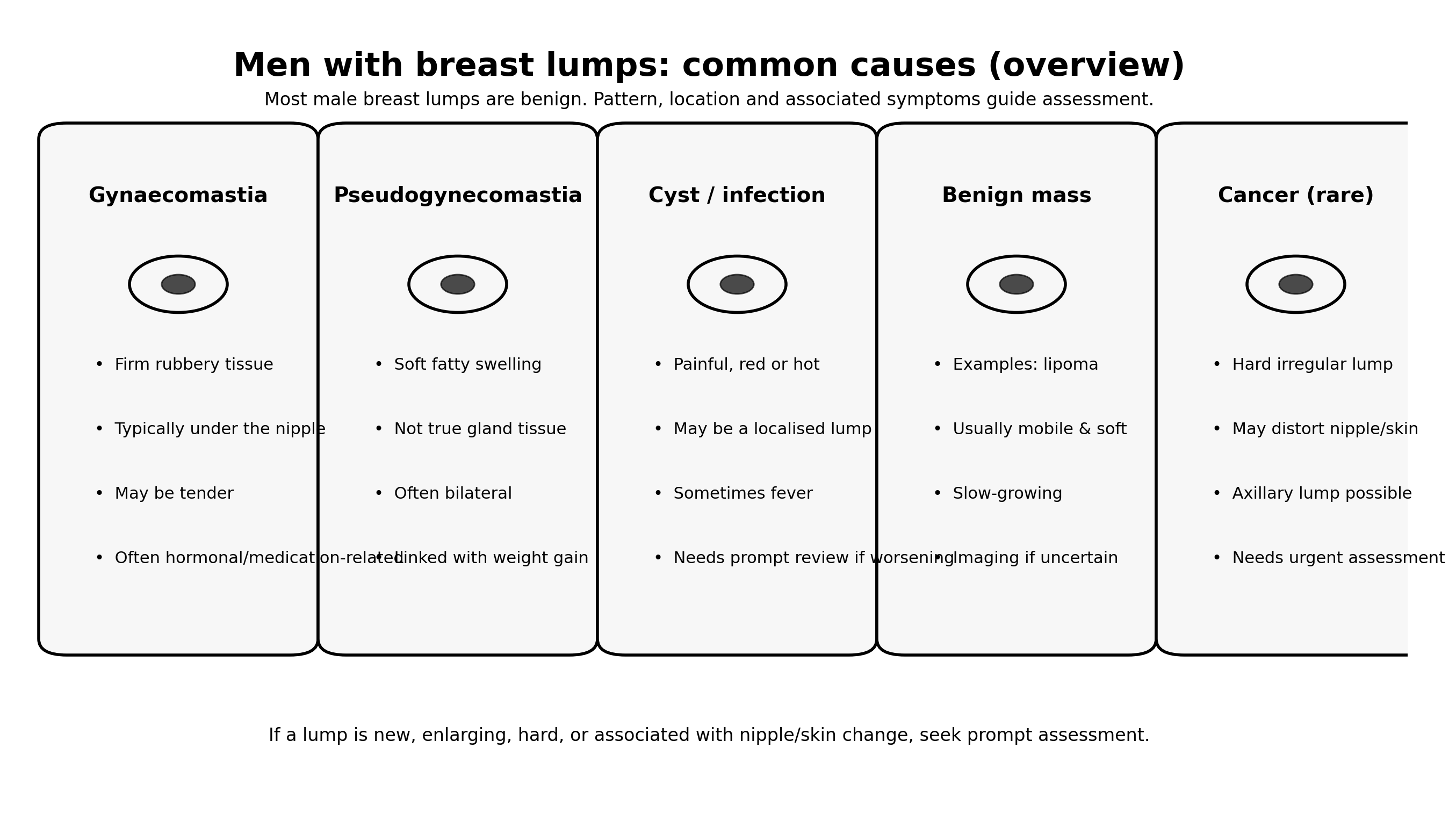
- Most male breast lumps are benign, commonly related to gynaecomastia (true gland tissue) or fatty swelling.
- Red flags include a hard irregular lump, nipple distortion/skin change, blood-stained discharge, or an armpit lump.
- Ultrasound is a targeted test that helps determine what the lump represents (for example gland tissue, cyst/infection, benign mass, or a suspicious lesion).
- If imaging identifies a suspicious finding, the pathway may include biopsy to confirm the diagnosis.
What counts as a “breast lump” in men?
Men can develop lumps or swelling in the breast area for several reasons. Some are within the breast tissue itself (especially under the nipple), and some are in the skin, fat, or chest wall structures. The clinical question I focus on is: Is this typical gynaecomastia, or is it a discrete indeterminate lump that needs imaging?
Common causes of breast lumps in men
1) Gynaecomastia (true gland tissue)
Gynaecomastia is the most common benign explanation for male breast swelling. It often feels like a firm or rubbery disc under the nipple and can be tender. It may be related to hormone changes, medications, liver disease, thyroid conditions, or simply occur without a clear trigger.
2) Pseudogynecomastia (fatty swelling)
This is not true gland growth. It is a soft enlargement due to fat distribution and is more likely with weight gain. It tends to feel more diffuse rather than a discrete subareolar lump.
3) Cyst or infection (abscess / inflamed skin lesion)
Painful redness, warmth, swelling, or a lump that feels “angry” can be due to an inflamed skin cyst, infection, or an abscess. If you have fever or rapidly worsening symptoms, you should seek prompt clinical review.
4) Benign masses (for example lipoma)
Some benign lumps are slow-growing and mobile under the skin (for example lipomas). When the diagnosis is unclear clinically, ultrasound can usually characterise these confidently.
5) Male breast cancer (uncommon)
Male breast cancer is uncommon, but we take it seriously because it can present as a lump—often with additional features such as nipple distortion, skin tethering, or an armpit lump. This is why red flags matter.
When to seek assessment
I recommend assessment if your lump is new, enlarging, persistent, or if you are simply unsure what it is. Please seek prompt clinical assessment if you have any of the following red-flag features:

Seek prompt assessment if you have:
- A new lump that is hard, irregular, or fixed
- A lump that is enlarging or not settling
- Nipple distortion, new inversion, or eczema-like nipple change
- Blood-stained or spontaneous nipple discharge
- Skin dimpling, ulceration, or persistent redness
- A lump in the armpit (axillary lump)
- Signs of infection with systemic symptoms (fever/unwellness), especially if worsening
What happens at assessment?
A safe evaluation is usually structured as:
- History: onset, change over time, tenderness, medications, recent weight change, and any nipple/skin symptoms.
- Examination: is it subareolar gland tissue, a discrete mass, or something skin/chest-wall related? Is there an axillary lump?
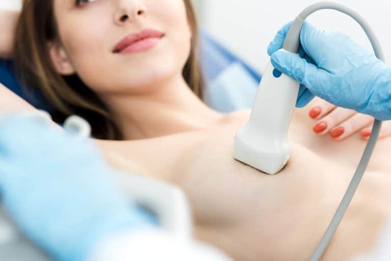
- Imaging (when indicated): targeted ultrasound (and sometimes mammography depending on age, features, and pathway).
How ultrasound supports symptom-led evaluation
Ultrasound is a very practical tool in men because it is targeted. We scan the exact area you are concerned about and answer the core diagnostic questions:
- Is the lump in the breast tissue, the skin, the fat, or deeper structures?
- Is it solid or cystic?
- Is there a typical subareolar gynaecomastia pattern?
- Are there features that look suspicious and need a biopsy pathway?
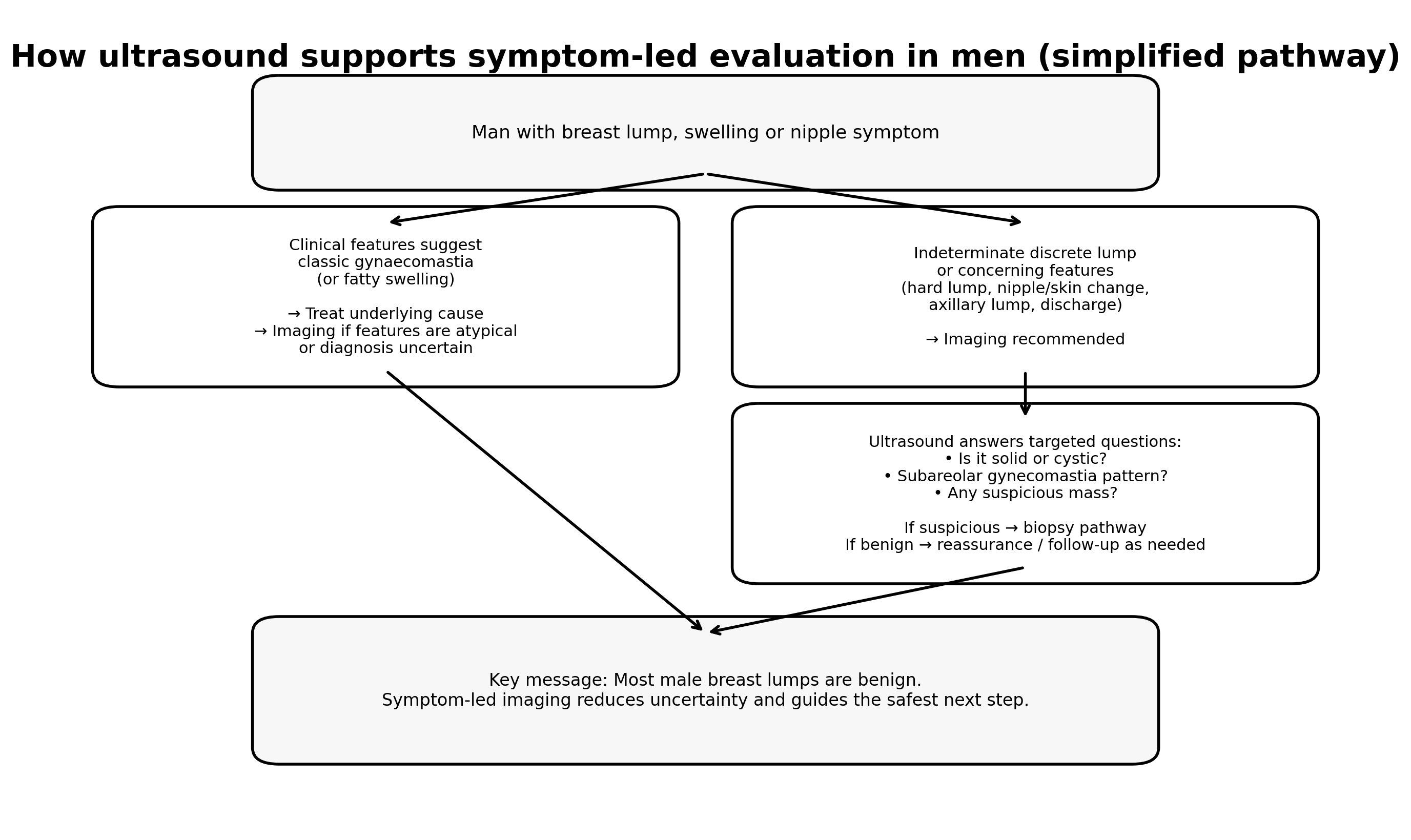
What an ultrasound may show
In many men, ultrasound confirms a benign explanation quickly. For example, gynaecomastia has a recognisable appearance beneath the nipple, whereas a discrete mass is assessed carefully and measured, with Doppler used when appropriate.

If the scan is normal or benign
If imaging confirms a benign cause, the next step is typically reassurance and guidance. Depending on the clinical scenario, I may advise follow-up with your GP to consider medication review, hormonal assessment, or monitoring over time.
If something needs further investigation
If ultrasound identifies a suspicious or indeterminate lesion, the safest next step is usually to follow a standard diagnostic pathway, which may include mammography and/or a biopsy referral. The goal is to avoid uncertainty and reach a definite diagnosis.
FAQs
Is a painful lump more likely to be benign?
Pain is common with benign causes (including gynaecomastia or infection), but pain alone does not rule out significant pathology. The overall pattern and examination findings determine whether imaging is recommended.
Can gynaecomastia be on one side only?
Yes. It can be unilateral or bilateral. The typical location is directly under the nipple, and it may be tender.
Do men ever need mammography?
Sometimes. In men with a discrete indeterminate lump or suspicious clinical features, mammography can be used alongside ultrasound depending on age and pathway. The imaging plan is individualised.
What should I do while waiting for assessment?
Avoid repeatedly squeezing or checking the area aggressively, as this can worsen tenderness. If you develop red flags (nipple/skin changes, discharge, armpit lump, rapid growth, fever), seek prompt clinical review.
Booking note (IUS London)
If you have a new breast lump or nipple symptom, a symptom-led ultrasound assessment can help clarify the cause and guide the safest next step. If there are red flags, I recommend prompt clinical assessment.
Next step: Contact IUS London to book