Follicle Size by Cycle Day — What\'s Normal: A Comprehensive Guide to Ovarian Follicle Development and Fertility
Understanding the intricate journey of ovarian follicle development is paramount for anyone navigating the complexities of fertility, whether through natural conception or assisted reproductive technologies (ART). Follicles, the tiny, fluid-filled sacs nestled within the ovaries, serve as the nurseries for immature eggs (oocytes). Their growth, maturation, and eventual release are meticulously orchestrated by a symphony of hormones, culminating in the pivotal event of ovulation.
This guide delves into the fascinating biology of ovarian follicles, offering a detailed exploration of their anatomy, physiology, and the hormonal mechanisms that govern their progression. We will also provide an in-depth look at the patient experience during follicle tracking scans, present anonymised clinical scenarios from our practice, and address frequently asked questions to empower you with knowledge about this critical aspect of reproductive health.
The Ovarian Follicle: A Deeper Dive into Anatomy and Physiology
At birth, a female\'s ovaries are endowed with a finite reserve of primordial follicles, each containing an oocyte arrested in the prophase I stage of meiosis. This dormant pool represents the entirety of a woman\'s reproductive potential. Throughout her reproductive lifespan, a select cohort of these primordial follicles is continuously recruited in a cyclical manner to embark on a journey of growth and differentiation, a process known as folliculogenesis.
Folliculogenesis is a highly complex and tightly regulated biological cascade involving cellular proliferation, intricate cell-to-cell communication, and precise hormonal signaling. It is a continuous process, with follicles at various stages of development coexisting within the ovary at any given time.
Stages of Follicle Development: From Primordial to Preovulatory
1. Primordial Follicles: These are the most rudimentary follicular units, characterized by a quiescent oocyte enveloped by a single layer of flattened, squamous granulosa cells. They represent the ovarian reserve and remain in a state of suspended animation until activated.
2. Primary Follicles: Upon recruitment from the primordial pool, the flattened granulosa cells transform into a cuboidal shape and begin to proliferate, forming multiple layers around the oocyte. Concurrently, a crucial extracellular matrix, the zona pellucida, starts to form, encasing the oocyte. This glycoprotein layer plays a vital role in sperm binding and fertilization.
3. Secondary Follicles: As granulosa cell proliferation continues, the surrounding ovarian stromal cells begin to differentiate, forming two distinct layers: the theca interna and the theca externa. The theca interna cells are rich in luteinizing hormone (LH) receptors and are responsible for synthesizing androgens (male hormones) under LH stimulation. These androgens are then transported to the granulosa cells, which, under the influence of follicle-stimulating hormone (FSH), possess the enzyme aromatase to convert them into estrogens (female hormones). The theca externa consists of connective tissue and smooth muscle cells, providing structural support to the growing follicle.
4. Antral (Tertiary) Follicles: A hallmark of this stage is the formation of a fluid-filled cavity within the granulosa cell layers, known as the antrum. The follicular fluid within the antrum contains hormones, growth factors, and other substances essential for oocyte maturation. The emergence of the antrum signifies a critical shift in follicular development, as these follicles become increasingly dependent on Follicle-Stimulating Hormone (FSH) for their continued growth and survival. It is at the antral stage that follicles become large enough to be visualized and measured using ultrasound imaging.
5. Graafian (Preovulatory) Follicle: In a typical menstrual cycle, out of the cohort of antral follicles that begin to develop, only one—the dominant follicle—is selected to continue its rapid growth towards ovulation. This selection process is driven by the dominant follicle\'s enhanced sensitivity to FSH. The Graafian follicle can reach a substantial size, typically between 18-24mm in diameter, just prior to ovulation. The remaining follicles in the cohort, unable to compete for the dwindling FSH supply, undergo a process of degeneration called atresia.
Hormonal Regulation: The Endocrine Symphony of Folliculogenesis
The growth and development of ovarian follicles are meticulously regulated by a complex interplay of hormones, primarily originating from the hypothalamus, pituitary gland, and the ovaries themselves:
- Follicle-Stimulating Hormone (FSH): Secreted by the anterior pituitary gland, FSH is the primary driver of follicular growth. It stimulates the proliferation of granulosa cells and induces the expression of aromatase, facilitating estrogen production.
- Luteinizing Hormone (LH): Also secreted by the anterior pituitary, LH plays a crucial role in androgen production by the theca interna cells and, most importantly, triggers the final maturation of the oocyte and ovulation through a surge in its levels.
- Estrogen: Produced by the granulosa cells, estrogen exerts both positive and negative feedback effects. Initially, rising estrogen levels inhibit FSH secretion, contributing to dominant follicle selection. However, a sustained high level of estrogen triggers the LH surge, leading to ovulation.
- Progesterone: Primarily produced by the corpus luteum (the remnant of the ovulated follicle), progesterone prepares the uterus for pregnancy and inhibits further follicular development.
This intricate hormonal feedback loop ensures that typically only one dominant follicle matures and ovulates each cycle, preventing multiple pregnancies in natural cycles.
Follicle Tracking: The Patient Experience During a Scan
Follicle tracking, most commonly performed via transvaginal ultrasound (TVS), is an indispensable tool in modern fertility management. It provides real-time visual information about the number, size, and growth rate of ovarian follicles, allowing clinicians to predict ovulation, optimize the timing of intercourse or intrauterine insemination (IUI), and guide ovarian stimulation protocols for in vitro fertilization (IVF).
Preparing for Your Scan: What to Expect
Patients are generally provided with specific instructions prior to their appointment. For transvaginal ultrasounds, a comfortably empty bladder is usually preferred, as a full bladder can sometimes obscure the view of the ovaries. There are typically no dietary restrictions. It is always advisable to communicate any anxieties or discomfort you may anticipate to the sonographer or clinician, who can provide reassurance and adjust the procedure as needed. Open communication ensures a more comfortable and effective experience.
The Scan Procedure: A Detailed Walk-Through
The transvaginal ultrasound involves the gentle insertion of a thin, sterile, and lubricated ultrasound probe into the vagina. This probe is specially designed to emit high-frequency sound waves that penetrate the pelvic organs. These sound waves then bounce off internal structures, such as the ovaries, uterus, and fallopian tubes, and are converted into detailed, real-time images displayed on a monitor. The sonographer will systematically maneuver the probe to obtain comprehensive views of both ovaries, meticulously measuring the diameter of all visible follicles. They will also assess the thickness and appearance of the endometrial lining, which is crucial for embryo implantation.
The procedure is generally well-tolerated, with most patients experiencing only mild pressure or a sensation of fullness. The entire scan typically lasts between 10 to 20 minutes. Throughout the scan, the sonographer may explain what they are observing on the screen, providing a sense of involvement and understanding for the patient.
Post-Scan Consultation and Next Steps
Immediately following the scan, the clinician will review the ultrasound images and discuss the findings with you. This consultation is an opportunity to understand:
- Follicle Count and Size: The number of developing follicles and their individual measurements.
- Endometrial Thickness: The receptivity of the uterine lining.
- Hormone Levels: Often, blood tests for hormones like estradiol and LH are performed concurrently with ultrasound scans to provide a complete picture of follicular activity.
- Implications for Your Cycle/Treatment: Based on these observations, the clinician will explain what the findings mean for your natural cycle or your specific fertility treatment plan. This might include predicting the optimal window for ovulation, adjusting medication dosages in stimulated cycles, or scheduling further monitoring scans.
This comprehensive approach ensures that decisions regarding your fertility journey are informed and tailored to your individual physiological responses.
Normal Follicle Growth and Ovulation: Key Milestones
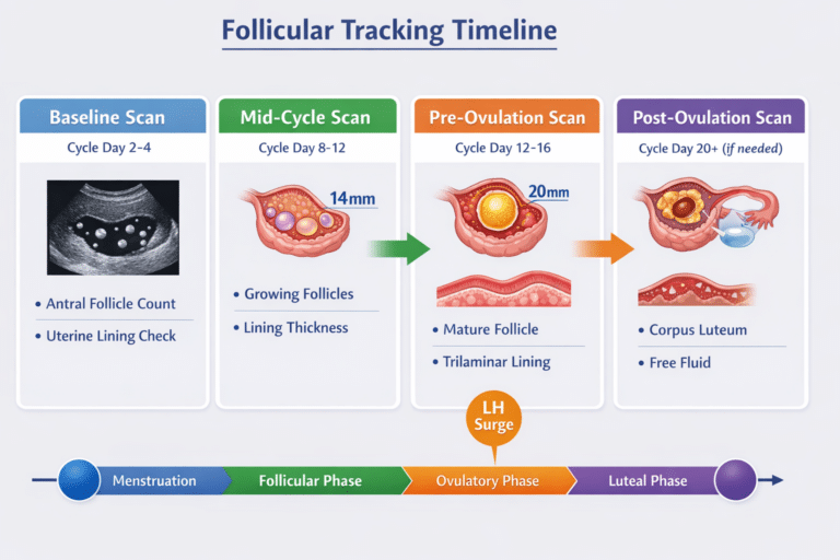
In a natural, unstimulated menstrual cycle, the dominant follicle typically grows at an average rate of 1-2mm per day during the follicular phase. This consistent growth rate is a key indicator of healthy follicular development. The optimal size for a mature, ovulatory follicle is generally considered to be between 18-24mm just before the LH surge triggers its release. Follicles within this range are most likely to contain a mature, viable egg capable of fertilization.
Basal Antral Follicle Count (AFC): Assessing Ovarian Reserve
An Antral Follicle Count (AFC) is a cornerstone of fertility assessment, usually performed early in the menstrual cycle (typically between Day 2 and Day 5). This ultrasound-based assessment involves counting the number of small antral follicles (measuring 2-10mm in diameter) present in both ovaries. The AFC is a highly reliable surrogate marker for ovarian reserve, providing a quantitative estimate of the remaining egg supply. A higher AFC generally correlates with a better response to ovarian stimulation and a greater chance of success in fertility treatments. It helps clinicians tailor stimulation protocols and manage patient expectations.
Follicle Monitoring in Stimulated Cycles: Maximizing Outcomes
In assisted reproductive technologies such as In Vitro Fertilization (IVF), the primary objective is to stimulate the ovaries to produce multiple mature follicles. This is achieved through controlled ovarian hyperstimulation using various fertility medications. During these stimulated cycles, meticulous follicle tracking is even more critical. Clinicians closely monitor the growth of multiple lead follicles, aiming for a similar size range (typically 18-22mm) before administering a trigger shot (e.g., hCG) to induce final maturation and ovulation or scheduling egg retrieval. The precise timing of the trigger is crucial for maximizing the yield of mature, high-quality eggs.
Regular ultrasound monitoring during stimulated cycles allows for dynamic adjustments to medication dosages and timing, minimizing risks such as Ovarian Hyperstimulation Syndrome (OHSS) and optimizing treatment efficacy. The goal is to achieve synchronous growth of a sufficient number of follicles, each containing a viable oocyte.
From Our Practice: Anonymised Clinical Scenarios and Insights
At IUS London, we encounter a diverse range of patient profiles, each presenting unique challenges and opportunities in their fertility journey. Here, we share anonymised clinical scenarios to illustrate the practical application of follicle tracking and the nuanced decision-making involved.
Case Study 1: Sarah – The Optimistic First-Timer with PCOS Tendencies
Sarah, a 32-year-old professional, presented with a history of irregular menstrual cycles and had been trying to conceive for over a year without success. Her initial diagnostic workup, including hormonal assays and a basal antral follicle count (AFC), revealed an AFC of 12, suggesting a healthy ovarian reserve, but also indicated some polycystic ovary syndrome (PCOS) tendencies due to elevated AMH levels and a history of anovulation. Given her age and good ovarian reserve, we initiated a mild ovarian stimulation protocol for her first IVF cycle.
During her monitoring scans, we observed a promising response. By day 9 of stimulation, Sarah had three dominant follicles measuring 16mm, 17mm, and 18mm, alongside several smaller follicles that were actively growing. To allow these smaller follicles to catch up and to ensure optimal maturity of the lead follicles, we continued stimulation for an additional two days. On day 11, her lead follicles had reached ideal sizes of 19mm, 20mm, and 21mm. At this point, an hCG trigger shot was administered to induce final oocyte maturation. Precisely 36 hours later, we performed egg retrieval, successfully collecting 8 mature eggs. This case highlights the importance of individualized protocols and dynamic monitoring to achieve optimal outcomes, even in seemingly straightforward cases.
Case Study 2: Maria – The Challenging Responder with Diminished Ovarian Reserve
Maria, a 38-year-old patient, had a more complex fertility history, having undergone several unsuccessful IVF cycles at other clinics. Her basal AFC was significantly lower, with only 5 antral follicles, indicative of diminished ovarian reserve. Recognizing the need for a tailored approach, we designed a customized stimulation protocol aimed at maximizing the quality of the few available oocytes rather than focusing solely on quantity.
Follicle growth in Maria\'s cycle was initially slower than anticipated. By day 12 of stimulation, she had two follicles measuring 17mm and 18mm. Given her history and the importance of each follicle, we extended the stimulation by one day to allow for further maturation. On day 13, these follicles measured 18.5mm and 19.5mm, respectively. Despite the lower number of mature follicles, we proceeded with the hCG trigger. The subsequent egg retrieval yielded 2 high-quality mature eggs. Crucially, one of these eggs resulted in a successful pregnancy, underscoring that even with diminished ovarian reserve, a personalized and carefully managed approach can lead to positive outcomes.
Case Study 3: Eleanor – The Unexpected Early Responder
Eleanor, 30, was undergoing her first IVF cycle due to male factor infertility. Her AFC was excellent, and she was expected to be a good responder. However, during her initial monitoring, we noticed a rapid growth of follicles, with several reaching 18mm by day 7 of stimulation. This early response, while positive in terms of follicle development, posed a risk of premature ovulation or OHSS if not managed carefully. We immediately adjusted her medication dosage downwards and introduced a GnRH antagonist to prevent an LH surge. By day 9, her follicles were optimally sized between 19-21mm, and we proceeded with a GnRH agonist trigger, followed by egg retrieval. This case illustrates the need for vigilant monitoring and flexible protocol adjustments to prevent complications and ensure successful egg retrieval.
Comprehensive Frequently Asked Questions (FAQs) and Common Patient Concerns
Navigating fertility treatments can be overwhelming, and patients often have numerous questions and concerns. Here, we address some of the most common queries regarding follicle size and development.
Q1: What if my follicles are growing too slowly or too quickly?
Slow Follicle Growth: If follicles are growing slower than the expected 1-2mm per day, it can indicate a suboptimal response to the stimulation medication, or it may point to underlying ovarian issues. Your clinician will carefully evaluate your hormone levels (e.g., estradiol) in conjunction with ultrasound findings. Adjustments to medication dosages, changes in the type of gonadotropin used, or extending the stimulation period are common strategies. In some cases, a different protocol might be considered for future cycles.
Rapid Follicle Growth: Conversely, very rapid follicle growth can sometimes lead to premature ovulation, where eggs are released before they are fully mature, or increase the risk of Ovarian Hyperstimulation Syndrome (OHSS). In such scenarios, medication dosages may be reduced, or an antagonist medication might be introduced to prevent an untimely LH surge. Close monitoring is key to managing both extremes of growth rate.
Q2: Can follicles be too large or too small for egg retrieval?
Follicles That Are Too Large: While larger follicles generally signify maturity, excessively large follicles (e.g., typically >25mm) may be post-mature or cystic. Post-mature eggs may have compromised quality, while cystic structures may not contain a viable egg. Your clinician will interpret the significance of large follicles in the context of your overall hormonal profile and the growth trajectory of other follicles.
Follicles That Are Too Small: Follicles that remain too small (e.g., <14-16mm) at the time of trigger or retrieval are less likely to contain a mature egg. Immature eggs cannot be fertilized. The goal of ovarian stimulation is to encourage as many follicles as possible to reach the optimal mature size range.
Q3: Does follicle size directly correlate with egg quality?
Follicle size is an important indicator of maturity, but it does not guarantee egg quality. A follicle within the optimal size range (18-24mm) is more likely to contain a mature, high-quality egg. However, egg quality is a complex factor influenced by numerous variables, including maternal age, genetics, lifestyle, and overall ovarian health. It is possible to have appropriately sized follicles that yield eggs of suboptimal quality, and conversely, occasionally smaller follicles can yield good quality eggs. Therefore, clinicians consider follicle size alongside other parameters, such as estradiol levels and patient history, to assess the overall likelihood of retrieving viable eggs.
Q4: Is it normal to have follicles of different sizes during a stimulated cycle?
Yes, it is very common and, to some extent, expected to have follicles of varying sizes during both natural and stimulated cycles. In a natural cycle, typically only one follicle becomes dominant, while the others undergo atresia. In stimulated cycles, the aim is to recruit and grow multiple follicles, but they rarely grow at precisely the same rate. The clinician\'s skill lies in timing the trigger shot when the majority of the lead follicles are within the optimal mature size range, balancing the maturity of larger follicles with the potential for smaller ones to catch up.
Q5: What is the significance of the endometrial lining thickness?
The endometrial lining, or the lining of the uterus, plays a critical role in successful embryo implantation. Its thickness and appearance are closely monitored during follicle tracking. An optimal endometrial thickness (typically 8-14mm) with a trilaminar (three-layered) appearance is generally considered favorable for implantation. If the lining is too thin or has an unfavorable appearance, it can reduce the chances of pregnancy, even if follicle development is optimal. Hormonal support may be provided to improve endometrial receptivity.
Q6: How many follicles are considered ideal for IVF?
There isn\'t a single \'ideal\' number, as it varies based on individual patient factors like age, ovarian reserve, and diagnosis. However, a good response in IVF typically involves retrieving 8-15 mature eggs. This range offers a good balance between maximizing the chances of finding a healthy embryo and minimizing the risk of OHSS. Retrieving too few eggs can limit embryo selection, while an excessive number might increase OHSS risk. The focus is always on quality over quantity, aiming for a reasonable number of mature, high-quality oocytes.
Q7: What is Ovarian Hyperstimulation Syndrome (OHSS) and how is it related to follicle size?
Ovarian Hyperstimulation Syndrome (OHSS) is a potentially serious complication of fertility treatments, particularly those involving ovarian stimulation. It occurs when the ovaries overreact to fertility medications, producing too many follicles and releasing excessive amounts of hormones. This can lead to fluid shifting from blood vessels into the abdominal cavity and, in severe cases, into the lungs. Symptoms range from mild (bloating, abdominal discomfort) to severe (severe abdominal pain, nausea, vomiting, shortness of breath, blood clots, kidney failure). The risk of OHSS is directly related to the number and size of developing follicles; a high number of large follicles increases the risk. Close monitoring of follicle growth and estradiol levels, along with judicious use of trigger medications and preventative strategies, are crucial to minimize OHSS risk.
Red Flags: When to Seek Immediate Medical Attention
While follicle tracking is a routine and generally safe procedure, certain symptoms or observations during fertility treatment warrant immediate medical attention. Recognizing these red flags can prevent complications and ensure timely intervention:
- No or minimal follicle growth despite stimulation: This could indicate a poor or absent response to ovarian stimulation, suggesting underlying issues with ovarian function or an inappropriate treatment protocol. It requires prompt re-evaluation by your fertility specialist.
- Premature ovulation: If ovulation occurs unexpectedly before the follicles have reached their optimal mature size, it can result in the release of immature eggs, significantly reducing the chances of successful fertilization. Symptoms might include sudden pelvic pain or a rapid drop in estradiol levels.
- Symptoms of Ovarian Hyperstimulation Syndrome (OHSS): As discussed, OHSS can range from mild to severe. Any experience of severe abdominal pain, significant bloating, persistent nausea or vomiting, sudden weight gain, decreased urination, or shortness of breath during or after ovarian stimulation cycles should be reported to your clinic immediately. Early detection and management are crucial.
- Sudden, severe pelvic pain: While mild discomfort is common, sharp, incapacitating pelvic pain could indicate ovarian torsion (twisting of the ovary), a rare but serious complication that requires emergency medical intervention.
- Unexplained fever or chills: These symptoms, especially after an egg retrieval procedure, could be indicative of an infection and require immediate medical assessment.
Always err on the side of caution and contact your fertility team if you experience any concerning symptoms during your treatment journey.
Call to Action: Partner with IUS London for Your Fertility Journey
Navigating the complexities of fertility requires expert guidance, personalized care, and a deep understanding of reproductive science. If you have concerns about your fertility, are considering fertility treatments, or simply wish to gain a clearer understanding of your reproductive health, the specialist team at IUS London is here to support you.
Our clinic offers advanced diagnostic services, modern follicle tracking, and individualized treatment plans tailored to your unique needs and circumstances. We are committed to providing compassionate, evidence-based care to help you achieve your family-building goals.
Contact IUS London today to schedule a comprehensive consultation. Let us partner with you on your path to parenthood, providing clarity, support, and the highest standard of medical expertise. Your journey to understanding and optimizing your fertility begins here.
Book Your Private Ultrasound Scan in London
Same-day appointments available in Kensington. No GP referral required. HCPC-registered consultant sonographers.