Ultrasound and Fertility — What a Scan Can and Cannot Show
A scan cannot tell you whether you are infertile. What it can do is answer several of the structural questions a fertility investigation asks — and rule things out that would otherwise stay unknown.
The question people arrive with is usually some version of “can an ultrasound show if I am infertile?” — so it deserves answering first, plainly. No, it cannot. Fertility is not a structure that can be imaged. It depends on egg quality, sperm, hormones, whether the fallopian tubes are open, and whether an embryo implants — and ultrasound sees almost none of that.
What it does is different and still genuinely useful: it examines the anatomy involved, counts what can be counted, and identifies things that are treatable. A normal fertility scan does not mean you will conceive easily. An abnormal one does not mean you will not. What either gives you is information a fertility specialist can act on.
What ultrasound sees — and what it does not
| Ultrasound can assess | Ultrasound cannot assess |
|---|---|
| Antral follicle count — an indicator of ovarian reserve | Egg quality, which matters more than number and cannot be imaged at all |
| The shape and cavity of the uterus | Whether the fallopian tubes are open — normal tubes are invisible on ordinary ultrasound |
| Fibroids and polyps, and whether they distort the cavity | Sperm — a separate investigation entirely |
| Endometrial thickness and pattern | Whether an embryo will implant |
| Polycystic ovarian appearance | Chromosomes or genetics |
| Endometriomas, and fluid-filled tubes suggesting blockage | Hormone levels — these are blood tests |
| Whether and when you ovulate, tracked across a cycle | Unexplained infertility, which by definition has no visible cause |
The tubal point is worth underlining, because it surprises people. Healthy fallopian tubes cannot be seen on a standard scan — they only become visible when abnormally distended with fluid. Testing whether tubes are open needs a dedicated test using contrast, such as HyCoSy or an HSG, arranged through a fertility clinic. A normal pelvic ultrasound says nothing either way about tubal patency.
You cannot see an egg on a scan
People search for this often, so: an egg is microscopic — around a tenth of a millimetre — and no ultrasound can show one. Neither can a scan show fertilisation, or an embryo in the days after conception.
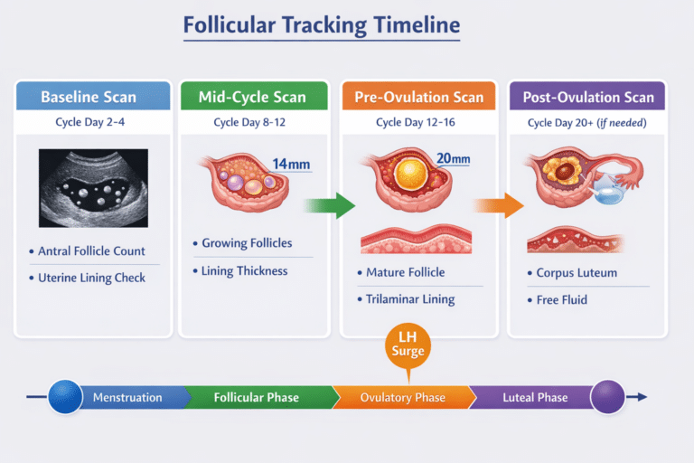
What is visible is the follicle: the fluid-filled sac an egg matures inside, which grows to roughly 18–24mm before releasing it. Follicle tracking watches those follicles grow and then disappear — and that disappearance is the evidence ovulation happened, not a sighting of the egg itself. Our guide to follicles versus cysts explains the distinction, which reports frequently blur.
After conception, the earliest thing visible is a gestational sac from around five weeks — weeks after fertilisation, not days.
Where a scan fits in a fertility investigation
A full investigation usually has four strands, and imaging is one of them:
- 1
Ovarian reserveAn antral follicle count by ultrasound, usually alongside an AMH blood test. The two together give a better picture than either alone — and both indicate quantity, not quality.
- 2
OvulationWhether you ovulate and when, assessed by follicle tracking across a cycle, sometimes with progesterone blood testing.
- 3
AnatomyA pelvic ultrasound assessing the uterus, cavity, endometrium and ovaries — identifying fibroids, polyps, endometriomas or a polycystic appearance.
- 4
Tubes and spermTubal patency testing and semen analysis. Neither is ultrasound, and neither is something we provide — both are arranged through a fertility clinic or your GP.
If you have been trying for a year — or six months if you are over 35 — the useful step is a GP appointment to begin that full investigation, rather than a scan on its own. A scan can start the picture, and we will tell you when what you actually need is the rest of it.
Ultrasound through an IVF or IUI cycle
This is where scanning does the most work, and where most fertility ultrasound actually happens — typically at your treating clinic as part of the cycle.
The baseline scan, early in the cycle before stimulation begins, checks the ovaries are quiet, counts antral follicles, and confirms there is no cyst that would complicate starting. Monitoring scans during stimulation then track how many follicles are developing and how fast, measured every few days alongside blood hormone levels. Clinicians are watching for enough follicles reaching maturity, growth at the expected rate, an endometrium thickening appropriately, and any sign of overstimulation.
Those measurements determine the trigger timing — when the final injection is given so eggs can be collected at the right moment. Egg collection itself is ultrasound-guided, with a needle passed under continuous imaging to reach each follicle, and embryo transfer is commonly guided by scanning too, to place the embryo accurately. A scan around two to three weeks after a positive test then confirms location and a heartbeat — our early pregnancy scan covers that stage.
Your questions answered
Can an ultrasound show if you are infertile?
Can a scan tell if my fallopian tubes are blocked?
Can you see an egg on an ultrasound?
What is an antral follicle count?
What monitoring happens during IVF stimulation?
Is a fertility scan painful?
When should I see a doctor about fertility?
What we offer
Ovarian reserve indicated by counting resting follicles in both ovaries.
About the scan →
£159Follicle TrackingWatching a dominant follicle grow, and confirming ovulation happened.
About the scan →
GuideReading Your Tracking ResultsWhat the follicle measurements on your report actually mean.
Read the guide →
Where a scan genuinely helps
Ovarian reserve and ovulation assessed by HCPC-registered sonographers, explained on screen, with a written report usually within two hours — and an honest word when what you need is the part of the investigation we do not provide.
Kensington, London W8 4ED
3 mins from Notting Hill Gate
This article is general information, not individual medical advice. Fertility investigation involves tests beyond ultrasound — including hormone bloods, tubal patency testing and semen analysis — which we do not provide; your GP or a fertility clinic can arrange them. Treatment monitoring described here is performed by your treating fertility clinic as part of your cycle.
Written and clinically reviewed by the HCPC-registered sonographers who perform this scan at IUS London — a CQC-registered diagnostic ultrasound clinic (Provider ID 1-2775844974). Your own findings are explained to you at the scan and set out in your report.
Author: Yianni Kiromitis, Senior Sonographer, HCPC RA38415
Medically reviewed: 4 August 2026