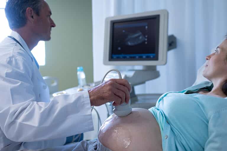
All mother to be are offered pregnancy scans resulting in pregnancy ultrasound being one of the most common ultrasound examinations in the United Kingdom.
When can you have a baby scan?
In our private clinic, we offer early pregnancy scans for as early as 5 weeks gestation, although we recommend having your first baby scan from 7 weeks unless you are concerned about miscarriage or an ectopic.
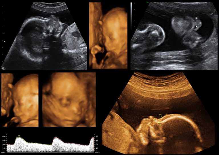
How many scans do you have during pregnancy?
Most healthy women receive two ultrasound scans during pregnancy. The first one is at 12 weeks, called a dating scan, and the second is at 18-22 weeks to confirm normal anatomy and possibly the sex of the baby.
Do you get an ultrasound at 6 weeks?
No. In the UK, you will not be offered a 6 weeks scan as a routine unless there are complications. If you would like a scan before 12 weeks for your peace of mind, you might have to opt-in for a private early scan.