Ultrasound technology plays a central role in understanding and managing fertility and reproductive health. For individuals navigating these aspects of their health, private ultrasound services offer accessible, personalized, and efficient care. This guide delves into how these scans work, their applications, and the advantages they offer.
What Are Fertility and Reproductive Health Ultrasound Scans?
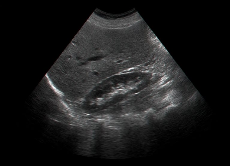
Ultrasound is a safe, non-invasive imaging technique that uses high-frequency sound waves to create detailed images of internal organs. In reproductive health, these scans are indispensable for visualizing the uterus, ovaries, fallopian tubes, and other pelvic structures. The two primary approaches are transabdominal scans, which involve a probe on the lower abdomen, and transvaginal scans, where a probe is inserted into the vaginal canal for a closer view of the pelvic organs.
These scans are critical in diagnosing conditions, monitoring treatments, and supporting fertility planning. For those pursuing private services, the benefits include shorter wait times, state-of-the-art equipment, and personalized care plans.
Key Uses of Ultrasound in Fertility and Reproductive Health
Fertility Assessments
Ultrasound scans provide essential insights into reproductive health. They are instrumental in monitoring ovarian follicle development, a key factor in ovulation. For individuals experiencing difficulties conceiving, scans can identify structural abnormalities such as uterine fibroids or polyps, which may hinder implantation.
Polycystic ovary syndrome (PCOS), a leading cause of infertility, is often diagnosed through ultrasound. The imaging reveals the characteristic "string of pearls" appearance of multiple small follicles in the ovaries. Research published in Fertility and Sterility highlights the accuracy of ultrasound in confirming PCOS, which affects up to 10% of reproductive-age women globally.
Saline contrast sonography, another ultrasound application, evaluates fallopian tube patency. This method involves introducing a saline solution into the uterus and tracking its flow to identify blockages. Studies in the Journal of Obstetrics and Gynaecology Research confirm this technique as less invasive yet highly effective compared to traditional dye tests.
Early Pregnancy Scans
Ultrasound is the gold standard for confirming pregnancy and determining gestational age. By visualizing the gestational sac and embryo, it establishes the viability of early pregnancy. The technology also identifies ectopic pregnancies, where the fertilized egg implants outside the uterus, a condition requiring immediate medical attention. Research in The Lancet underscores the importance of timely ultrasounds in reducing complications from ectopic pregnancies.
Assisted Reproductive Technology Support
For individuals undergoing treatments like in-vitro fertilization (IVF), ultrasounds are integral to the process. They monitor ovarian response to stimulation drugs, measure endometrial thickness, and guide procedures such as egg retrieval and embryo transfer. According to findings published in Reproductive BioMedicine Online, these scans significantly enhance the success rates of assisted reproductive technologies by ensuring precise timing and preparation.
Benefits of Private Ultrasound Scans for Fertility
Private services for fertility ultrasounds stand apart due to their emphasis on accessibility and customization. Waiting for a scan in a public healthcare setting can take weeks, delaying critical decisions in a fertility journey. Private providers, by contrast, often offer same-day appointments, ensuring timely access to care.
The quality of equipment in private clinics is another significant advantage. Many use advanced 3D and 4D imaging technologies, providing clearer and more detailed visuals compared to standard machines. Enhanced imaging leads to better diagnostic accuracy, as confirmed by research in the American Journal of Radiology.
Privacy and comfort also define the experience of private ultrasound appointments. Individuals can expect a calm and patient-focused environment where practitioners take the time to explain findings and address concerns.
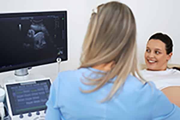
What to Expect During a Private Ultrasound Appointment
The process of undergoing a private ultrasound is straightforward and designed to prioritize patient comfort. Before the scan, instructions may include arriving with a full bladder for transabdominal scans to improve image quality. For transvaginal scans, no specific preparation is needed.
During the appointment, the sonographer applies a gel to the probe to facilitate sound wave transmission. For transabdominal scans, the probe is gently moved across the lower abdomen. Transvaginal scans involve inserting a thin probe into the vaginal canal, which typically causes minimal discomfort.
The entire procedure usually takes about 20-30 minutes. Results are often shared immediately, with detailed explanations provided by the clinician. Many private clinics also offer written reports and images for future reference.
Common Concerns and Questions
Is it safe to have multiple ultrasound scans?
Ultrasound is widely recognized as a safe imaging modality with no evidence of harm to patients. It uses sound waves, not radiation, making it ideal for repeated use during fertility treatments or pregnancy.
Will the scan be painful?
Most individuals report little to no discomfort during ultrasound scans. Transvaginal scans may feel slightly invasive, but the process is quick and performed with care to minimize unease.
What are the costs of private scans?
Prices for private ultrasounds vary depending on the provider, location, and type of scan. Comprehensive packages are often available, especially for individuals requiring multiple scans over time. Many clinics are transparent about fees, making it easier to plan expenses.
How to Choose a Private Ultrasound Clinic
Selecting the right clinic for private ultrasound services involves assessing several factors. Accreditation is paramount. Ensure the clinic and its sonographers are certified by recognized bodies such as the Care Quality Commission (CQC) in the UK.
The reputation of the clinic often reflects the quality of its services. Reviews and testimonials from previous clients can provide valuable insights. Look for feedback on professionalism, equipment quality, and clarity of communication.
State-of-the-art technology is another consideration. Clinics equipped with 3D and 4D ultrasound machines can provide enhanced diagnostic capabilities. Finally, consider practical aspects like the clinic's location and appointment flexibility, especially if frequent scans are needed.
Private ultrasound scans are an essential tool for understanding and managing fertility and reproductive health. Whether for diagnosing conditions, supporting treatments, or monitoring early pregnancies, they offer precise and timely insights that guide decisions and improve outcomes. By choosing private services, individuals gain access to high-quality care tailored to their needs.