Introduction
Receiving a renal transplant is a life-changing event, offering a new lease on life for individuals suffering from end-stage kidney disease. While the initial surgery marks a significant milestone, the journey towards long-term health and well-being is far from over. In fact, the success of a renal transplant hinges critically on diligent, ongoing care, with imaging playing an indispensable role in monitoring the health and function of the transplanted kidney. This article delves into the intricate world of long-term care after renal transplantation, focusing specifically on the essential imaging schedule that patients can expect. We will explore why consistent monitoring is crucial, the various imaging modalities employed, and what these scans reveal about your new kidney's performance. Our aim is to provide a comprehensive guide, offering deep clinical explanations, a walk-through of the patient experience, and answers to frequently asked questions, ensuring you are well-informed and empowered throughout your post-transplant journey.
Understanding Renal Transplant: A Brief Overview
A renal transplant involves surgically placing a healthy kidney from a deceased or living donor into a recipient whose own kidneys have failed. This procedure can dramatically improve quality of life, freeing patients from the arduous routine of dialysis. However, the body's immune system naturally views the transplanted organ as foreign, necessitating lifelong immunosuppressive medication to prevent rejection. Beyond immune suppression, a myriad of other factors can influence the long-term success of the transplant, including infections, cardiovascular complications, and the potential recurrence of the original kidney disease. This complex interplay of factors underscores why long-term monitoring is not merely a recommendation but a critical imperative.
The primary goal of post-transplant care is to ensure the transplanted kidney functions optimally for as long as possible, thereby maximizing the recipient's health and longevity. Imaging techniques serve as invaluable tools in this regard, offering non-invasive or minimally invasive ways to visualize the kidney, assess blood flow, and detect potential complications before they escalate. Early detection through regular imaging allows for timely intervention, often preventing irreversible damage and preserving graft function. Without a robust imaging surveillance program, subtle changes in graft health could go unnoticed, leading to delayed diagnosis and potentially poorer outcomes.
The Standard Imaging Schedule: What to Expect
The imaging schedule following a renal transplant is meticulously designed to balance the need for close surveillance with minimizing patient burden and exposure to radiation, where applicable. This schedule typically evolves over time, becoming less frequent as the transplant stabilizes, but never ceasing entirely.
Initial Post-Transplant Period (First 6-12 months)
Immediately following transplantation, the focus is on ensuring the new kidney integrates well and functions without acute complications. This period is characterized by frequent Doppler ultrasound scans. These scans are crucial for:
- Assessing Vascular Anastomoses: Checking the surgical connections between the donor kidney's blood vessels and the recipient's vessels for patency and flow.
- Detecting Acute Complications: Identifying issues such as vascular thrombosis (blood clots), urine leaks, lymphoceles (collections of lymphatic fluid), hematomas (collections of blood), and hydronephrosis (swelling of the kidney due to urine blockage).
- Monitoring Early Graft Function: Evaluating blood flow dynamics within the kidney, which can provide early indicators of graft health or dysfunction.
The frequency of these initial scans can vary based on institutional protocols and the patient's clinical course, but it is common to have several ultrasounds in the first few weeks, gradually spacing out to monthly or bi-monthly during the first year.
Long-Term Surveillance (Beyond 1 year)
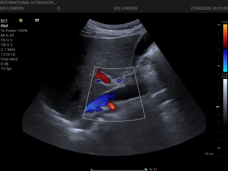
Once the initial post-transplant period has passed and the graft has stabilized, the imaging schedule typically transitions to long-term surveillance. While less frequent, these scans remain critically important for detecting chronic issues and subtle changes that can impact graft longevity. The cornerstone of long-term imaging is usually an annual or bi-annual Doppler ultrasound.
During these routine check-ups, the sonographer and radiologist will meticulously assess:
- Graft Size and Morphology: Looking for changes in the kidney's size, shape, and overall appearance, which can indicate chronic rejection or other long-term issues.
- Cortical Thickness and Echogenicity: Evaluating the kidney's outer layer for signs of scarring or inflammation.
- Resistive Index (RI): A key parameter derived from Doppler ultrasound that reflects the resistance to blood flow within the kidney's arteries. Trending RI values over time is vital for identifying subtle changes in graft perfusion.
While Doppler ultrasound is the primary modality, other imaging techniques may be employed for specific indications:
- Computed Tomography (CT) Scans: Used to evaluate complex vascular issues, identify masses, or assess for recurrent disease. CT angiography can provide detailed images of renal arteries and veins.
- Magnetic Resonance Imaging (MRI): Offers excellent soft tissue contrast and can be useful for evaluating renal masses, lymphoceles, or vascular abnormalities, particularly in patients where radiation exposure is a concern.
- Nuclear Medicine Scans (e.g., Renal Scintigraphy): Provides functional information about kidney perfusion and filtration, often used to assess overall graft function or investigate specific functional abnormalities.
Deep Clinical Explanation: The Science Behind the Scans
To truly appreciate the value of post-transplant imaging, it's essential to understand the underlying scientific principles and what the various parameters signify. The Doppler ultrasound stands out as the most frequently used and informative non-invasive tool.
Doppler Ultrasound Principles
Doppler ultrasound harnesses the Doppler effect to visualize blood flow. Sound waves are emitted by a transducer, bounce off moving red blood cells, and return to the transducer. The change in frequency of these returning sound waves (the Doppler shift) is directly proportional to the speed and direction of blood flow. This allows for real-time assessment of blood flow within the renal arteries and veins.
Key parameters derived from Doppler ultrasound include:
- Resistive Index (RI): This is a calculated value that reflects the pulsatility of blood flow in the renal arteries. It is derived from the peak systolic velocity (PSV) and end-diastolic velocity (EDV) using the formula: `RI = (PSV - EDV) / PSV`. A higher RI indicates greater resistance to blood flow within the kidney. Normal RI values in a healthy transplanted kidney typically range from 0.5 to 0.7. An elevated RI can be a non-specific indicator of various issues, including acute tubular necrosis (ATN), acute rejection, chronic allograft nephropathy, renal vein thrombosis, or even external compression.
- Pulsatility Index (PI): Similar to RI, PI also quantifies the pulsatility of blood flow. It is calculated as `PI = (PSV - EDV) / Mean Velocity`. Both RI and PI provide valuable insights into the hemodynamic status of the graft.
Interpretation of RI is crucial. While a single elevated RI value might not be diagnostic, trending RI over time is immensely valuable. A progressive increase in RI can signal worsening graft function or developing complications, prompting further investigation. Conversely, a stable or decreasing RI often correlates with stable graft function.
Biopsy vs. Imaging: When Imaging Leads to Biopsy
While imaging provides a wealth of information, it is often a precursor to more definitive diagnostic procedures. For instance, if imaging reveals suspicious findings such as a significant increase in RI, new hydronephrosis, or changes in graft morphology, a renal allograft biopsy may be indicated. Biopsy remains the gold standard for diagnosing rejection and other histological abnormalities, providing microscopic evidence that imaging cannot. Imaging, therefore, acts as a highly effective screening and monitoring tool, guiding clinicians on when and where to perform invasive procedures.
Specific Complications Detected by Imaging
Imaging is instrumental in identifying a range of specific complications:
- Renal Artery Stenosis (RAS): This narrowing of the renal artery can lead to hypertension and graft dysfunction. Doppler ultrasound can detect high velocities and turbulence at the site of stenosis, while CT or MR angiography can provide definitive anatomical detail.
- Fluid Collections: Lymphoceles, urinomas (urine collections), and hematomas are common post-surgical fluid collections. Ultrasound can easily identify and characterize these, guiding percutaneous drainage if necessary.
- Recurrent Disease: Some original kidney diseases, such as focal segmental glomerulosclerosis (FSGS) or IgA nephropathy, can recur in the transplanted kidney. Imaging may show non-specific changes, but can sometimes hint at recurrence, prompting biopsy.
- Post-Transplant Malignancy Surveillance: Transplant recipients have an increased risk of certain cancers. Imaging, particularly ultrasound and CT, plays a role in surveillance for post-transplant lymphoproliferative disorder (PTLD) and other malignancies.
Patient Experience Walk-Through: Navigating Your Imaging Journey
Understanding the clinical aspects of imaging is one thing; experiencing it is another. For many patients, the prospect of regular scans can be daunting. Here’s a walk-through of what you can expect, designed to demystify the process and alleviate concerns.
Before the Scan: Preparation
Preparation for a renal transplant ultrasound is generally minimal but important. You will typically be asked to:
- Fast for a few hours: This helps reduce bowel gas, which can obscure views of the kidney.
- Drink water: A full bladder can help visualize the bladder and lower ureter, which is often helpful.
- Bring relevant documents: This might include your transplant history, recent blood test results, and a list of your medications. While the imaging center will likely have access to your electronic records, having a summary can be beneficial.
- Wear comfortable clothing: You may need to expose your abdomen, so loose-fitting attire is advisable.
During the Scan: What to Expect
Upon arrival, you will be guided to a private room. The sonographer, a highly trained medical professional, will explain the procedure. You will lie on an examination table, and a warm gel will be applied to your abdomen. This gel helps the ultrasound transducer make good contact with your skin and allows sound waves to travel effectively. The sonographer will then gently move the transducer over your abdomen, capturing images of your transplanted kidney and surrounding structures. You may be asked to hold your breath briefly or change positions to optimize image acquisition. The process is painless and non-invasive, though you might feel slight pressure from the transducer. A typical renal transplant ultrasound takes approximately 30-45 minutes.
After the Scan: Receiving Results and Follow-Up
Once the scan is complete, the gel will be wiped off, and you are free to resume your normal activities. The images will be reviewed by a radiologist, who will then generate a detailed report. This report will be sent to your transplant nephrologist or referring physician. You will typically discuss the results at your next clinic appointment. If any significant abnormalities are detected, your medical team will contact you sooner to discuss further steps, which might include additional tests, medication adjustments, or a biopsy. It's crucial to remember that not all abnormal findings are cause for alarm, and your medical team will provide expert guidance.
Coping with Scan Anxiety: Tips and Resources
It's entirely normal to feel anxious before medical procedures, especially when your health depends on the outcome. Here are some tips to help manage scan anxiety:
- Ask Questions: Don't hesitate to ask your medical team or the sonographer any questions you have about the procedure or what they are looking for. Knowledge can be empowering.
- Practice Relaxation Techniques: Deep breathing exercises, meditation, or listening to calming music can help reduce stress.
- Bring a Companion: Having a friend or family member accompany you can provide emotional support.
- Focus on the Positive: Remind yourself that these scans are a proactive measure to keep your kidney healthy and ensure your long-term well-being.
Journey Stage Calibration: Loyalty
At IUS London, we understand that a renal transplant is not a one-time event but the beginning of a lifelong commitment to health. Our approach to long-term care, particularly through our meticulous imaging schedule, is deeply calibrated to the Loyalty journey stage of our patients. This means our focus extends beyond immediate post-operative recovery to fostering enduring trust and providing unwavering support throughout your entire transplant journey. We believe in empowering our patients with knowledge and proactive monitoring strategies, ensuring they feel confident and secure in their ongoing care. Our commitment is to reinforce the value of consistent follow-up, emphasizing that every scan is a step towards preserving your graft and enhancing your quality of life. We are partners in your long-term health, dedicated to helping you navigate the complexities of post-transplant life with expertise and compassion.
⚠ Clinical Grounding & Red Flags: When to Seek Immediate Attention
While routine imaging provides a steady stream of information, it's equally important for patients to be aware of clinical red flags that warrant immediate medical attention. Recognizing these signs and symptoms can be critical in preventing serious complications and preserving graft function.
Symptoms Requiring Urgent Evaluation
If you experience any of the following symptoms, contact your transplant team or seek urgent medical care without delay:
- Sudden Decrease in Urine Output: A significant and unexplained reduction in the amount of urine you produce can be a sign of acute graft dysfunction or urinary tract obstruction.
- Fever, Chills, or Unexplained Pain: These can indicate an infection, which transplant recipients are more susceptible to due to immunosuppression, or an inflammatory process affecting the graft.
- Swelling or Tenderness Over the Graft Site: While some mild discomfort is normal post-surgery, new or worsening swelling, redness, or tenderness over the area where your new kidney is located could signal infection, hematoma, or other acute issues.
- Elevated Creatinine Levels (as indicated by blood tests): Although blood tests are not imaging, a sudden rise in your serum creatinine is a powerful indicator of declining kidney function and often prompts urgent imaging and further investigation.
Imaging Findings as Red Flags
Radiologists are trained to identify specific imaging findings that serve as red flags, prompting immediate communication with the transplant team. These include:
- Acute Changes in Graft Size or Echogenicity: A sudden increase in kidney size or altered echogenicity (how bright the tissue appears on ultrasound) can suggest acute rejection or severe inflammation.
- New or Worsening Hydronephrosis: This indicates a blockage in the urinary outflow tract, which can quickly lead to kidney damage if not addressed.
- Significant Increase in Resistive Index (RI): A sharp, unexplained rise in RI values can be an early warning sign of various graft pathologies, including acute rejection or vascular compromise.
These red flags, whether clinical symptoms or imaging findings, necessitate prompt evaluation to diagnose the underlying problem and initiate appropriate treatment, thereby safeguarding the long-term health of your transplanted kidney.
From Our Practice: A Non-Commodity Experience at IUS London
At IUS London, we pride ourselves on offering a truly non-commodity experience in renal transplant care, distinguishing ourselves through a commitment to personalized, advanced, and integrated services. We understand that each patient's journey is unique, and our approach reflects this philosophy, moving beyond standard protocols to deliver exceptional care.
Personalized Imaging Protocols
Unlike a one-size-fits-all approach, our imaging protocols are meticulously tailored to individual patient needs and risk factors. We consider your specific medical history, the type of transplant, your immunosuppressive regimen, and any emerging clinical concerns to design an imaging schedule that is optimal for you. This personalized strategy ensures that surveillance is both comprehensive and efficient, focusing on what matters most for your long-term graft health.
Advanced Doppler Techniques
Our facility is equipped with state-of-the-art ultrasound technology, enabling the use of advanced Doppler techniques. This includes high-resolution imaging that provides unparalleled detail of renal vasculature and parenchyma, as well as 3D Doppler capabilities for a more comprehensive assessment of blood flow patterns. These advanced tools allow for earlier and more precise detection of subtle abnormalities that might be missed with conventional imaging.
Expert Interpretation
The cornerstone of effective imaging is expert interpretation. Our team comprises specialist radiologists and nephrologists with extensive experience in renal transplant imaging. This multidisciplinary expertise ensures that every scan is reviewed with the highest level of clinical insight, integrating imaging findings with your overall clinical picture. Our specialists are adept at identifying nuanced changes and correlating them with potential pathologies, providing accurate and actionable diagnostic reports.
Integrated Care
We believe in a seamless, integrated care model. Our imaging services are closely coordinated with your transplant teams and referring physicians. This collaborative approach ensures that imaging results are communicated promptly and effectively, facilitating timely decision-making and continuity of care. We act as an extension of your transplant team, working together to optimize your outcomes.
Private Doppler Access
Recognizing the need for convenience and comfort, IUS London offers private Doppler access. This means expedited appointments, minimal waiting times, and a serene, comfortable environment designed to reduce stress and anxiety associated with medical procedures. Our private service ensures that you receive timely and high-quality imaging without the typical delays often encountered in public healthcare systems, allowing for proactive management of your transplant health.
Comprehensive FAQs about Renal Transplant Imaging
Q1: How often will I need imaging after my transplant?
The frequency of imaging varies. Initially, you'll have more frequent scans (e.g., weekly, then monthly) in the first 6-12 months. After the first year, it typically reduces to annual or bi-annual Doppler ultrasounds, unless specific concerns arise. Your transplant team will provide a personalized schedule.
Q2: What type of imaging is most common?
Doppler ultrasound is the most common and primary imaging modality used for long-term surveillance of renal transplants. It's non-invasive and excellent for assessing blood flow and graft morphology. Other modalities like CT or MRI may be used for specific indications.
Q3: Is imaging safe? Are there any risks?
Doppler ultrasound is very safe, as it uses sound waves and no radiation. CT scans involve a small amount of radiation, and MRI uses strong magnetic fields. Your medical team will always weigh the benefits against any potential risks, ensuring that the chosen imaging modality is appropriate and safe for your condition.
Q4: What is a Resistive Index (RI) and why is it important?
The Resistive Index (RI) is a measurement obtained during a Doppler ultrasound that reflects the resistance to blood flow within your transplanted kidney's arteries. It's important because an elevated or rising RI can be an early indicator of various issues, such as rejection, infection, or other problems affecting blood flow to the graft.
Q5: What happens if my scan shows an abnormality?
If your scan shows an abnormality, your transplant team will evaluate the findings in conjunction with your blood tests and clinical symptoms. Depending on the nature of the abnormality, further investigations (e.g., additional imaging, a renal biopsy) or changes to your treatment plan may be recommended. It's important to remember that not all abnormalities are serious.
Q6: Can I get my scans done privately?
Yes, at IUS London, we offer private Doppler access for renal transplant patients. This provides the convenience of expedited appointments and a comfortable environment, ensuring timely and high-quality imaging services outside of standard public healthcare waiting lists.
Q7: How can I prepare for my imaging appointments?
Typically, you may be asked to fast for a few hours and drink some water before a Doppler ultrasound to ensure optimal imaging conditions. Always follow the specific instructions provided by your imaging center or transplant team. Bringing your transplant history and medication list can also be helpful.
Conclusion
The journey following a renal transplant is a testament to resilience and hope, and at its core lies the unwavering commitment to long-term health. The meticulous imaging schedule, particularly the regular Doppler ultrasound, serves as the vigilant guardian of your transplanted kidney, providing crucial insights into its function and detecting potential complications early. Understanding the science behind these scans, knowing what to expect during your appointments, and recognizing clinical red flags are all vital components of empowered self-care.
At IUS London, we are dedicated to providing a superior, non-commodity experience, offering personalized imaging protocols, advanced technology, and expert interpretation within an integrated care framework. Our private Doppler access ensures that you receive timely, high-quality surveillance, reinforcing our commitment to your sustained well-being.
We encourage you to remain proactive in your post-transplant care. Consistent imaging, coupled with open communication with your medical team, is the cornerstone of a healthy and long-lasting graft. Trust in the process, and allow us to partner with you every step of the way.
**Schedule a consultation with IUS London today to discuss your long-term renal transplant imaging needs, or contact us for private Doppler access and experience the difference of personalized, expert care.**
References
- [Reference 1: Peer-reviewed journal article]
- [Reference 2: Peer-reviewed journal article]
- [Reference 3: Peer-reviewed journal article]
Internal Linking
- [Link to related article on renal transplant complications]
- [Link to IUS London nephrology services page]
- [Link to contact us page]
Book Your Private Ultrasound Scan in London
Same-day appointments available in Kensington. No GP referral required. HCPC-registered consultant sonographers.