Flank Pain and Kidney Stones — Which Test Finds Them?
Renal colic is one of the most severe pains there is. Here is what causes it, when it becomes an emergency, and an honest account of where ultrasound sits against CT.

Classic renal colic is unmistakable: severe pain starting in the loin and sweeping round towards the groin, coming in waves, with restlessness — people pace rather than lie still — and often nausea and blood in the urine. It happens when a stone lodges in the ureter and the kidney backs up behind it.
Stone pain with fever is an emergency
An obstructed kidney that becomes infected can deteriorate quickly and needs urgent hospital treatment. Go to A&E or call 999 if severe loin pain comes with fever or shaking chills, if you are vomiting and cannot keep fluids down, if you are passing little or no urine, or if you feel faint. Acute severe colic belongs in urgent care — not in a booked private appointment.
Not all flank pain is a stone
| What you notice | Commonly |
|---|---|
| Waves of severe loin-to-groin pain, restlessness, blood in urine | Ureteric stone |
| Loin pain with fever, burning and urinary frequency | Kidney infection — needs prompt treatment |
| Ache worse with movement or certain positions, tender to press on the back | Musculoskeletal |
| Sudden severe abdominal or back pain in an older adult with collapse | Aortic emergency — call 999 |
| One-sided pain with a known kidney cyst or previous stones | Often recurrence — still assess |
| Pelvic pain in women, sometimes mistaken for renal | Gynaecological — see our pelvic pain guide |
Ultrasound versus CT for stones
Being straight about this matters, because it is the one area where the test we sell is not the best test. A low-dose CT scan of the kidneys, ureters and bladder is the definitive test for stones — it finds them almost regardless of size or position, including small stones in the mid-ureter that ultrasound routinely misses. In acute colic, that is what an emergency department will do.
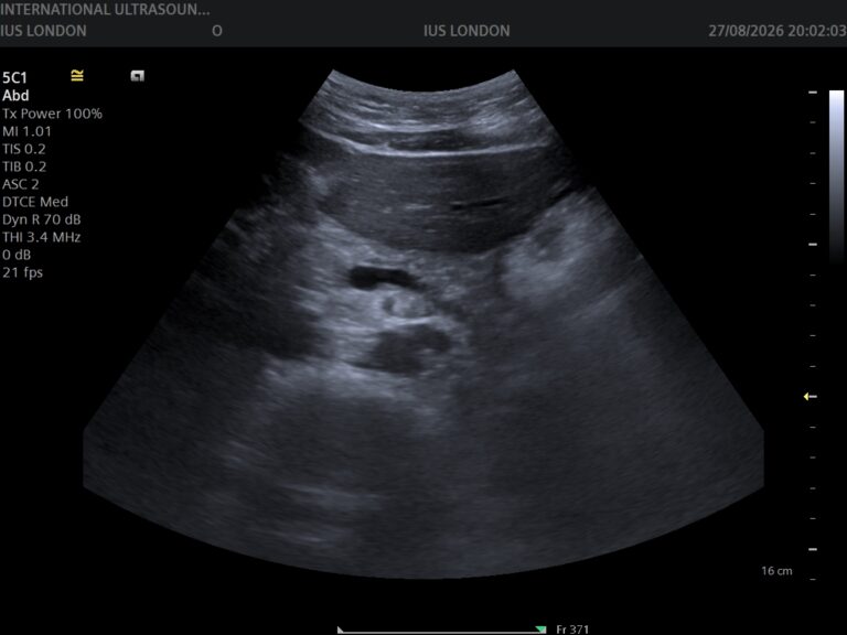
What ultrasound does well: it shows stones in the kidney itself, and — importantly — it shows hydronephrosis, the swelling of the collecting system that tells you a kidney is obstructed even when the stone itself cannot be seen. It also assesses kidney size and texture, finds cysts and masses, and looks at the bladder. It uses no radiation, which is why it is preferred in pregnancy and for repeated follow-up in people with recurrent stones.
So: acute severe colic goes to A&E, where CT is available. Ultrasound is well suited to non-acute assessment, to monitoring known stones over time, and to answering the kidney questions without another radiation dose. We do not offer CT.
When our scan is the right call
- 1
Not during an acute attackSevere colic, especially with fever or vomiting, is an A&E matter today. We will say so if you call us mid-attack.
- 2
Known stones being monitoredUltrasound follows stones over time without repeated radiation — a real advantage for anyone who forms them recurrently.
- 3
Settled or recurring milder painOnce the acute episode has passed, a scan assesses the kidneys, checks for obstruction, and looks for other causes.
- 4
The appointmentA comfortably full bladder helps. 30 minutes, no referral. Findings explained on screen as we scan, written report usually within two hours.
Your questions answered
Can an ultrasound see kidney stones?
Why choose ultrasound at all then?
How do I know if a stone has passed?
What is hydronephrosis?
How can I reduce the chance of more stones?
Do I need a full bladder for the scan?
Where to go from here
Once the acute episode has settled
Both kidneys, the collecting system and the bladder assessed without radiation — obstruction, stones, cysts and emptying — explained on screen as we scan, with the report usually within two hours.
Kensington, London W8 4ED
3 mins from Notting Hill Gate
This article is general information about flank pain and kidney stones, not individual medical advice. Severe loin pain with fever, vomiting, reduced urine output or faintness needs emergency care — see NHS guidance on kidney stones and kidney infection. Low-dose CT is the definitive test for stones; we do not offer CT.
Written and clinically reviewed by the HCPC-registered sonographers who perform this scan at IUS London — a CQC-registered diagnostic ultrasound clinic (Provider ID 1-2775844974). Your own findings are explained to you at the scan and set out in your report.
Author: Yianni Kiromitis, Senior Sonographer, HCPC RA38415 — over 20 years’ experience in NHS and private ultrasound
Medically reviewed: 9 August 2026