Fatty Liver — Symptoms, Causes, and Imaging

Fatty Liver: Understanding Symptoms, Causes, and Advanced Imaging at IUS London

Introduction

Fatty liver disease, medically known as hepatic steatosis, is a condition characterised by the accumulation of excess fat in the liver cells. While often benign in its early stages, it can progress to more severe forms, including inflammation (steatohepatitis), fibrosis, cirrhosis, and even liver failure or liver cancer. In the UK, the prevalence of fatty liver disease is rising, mirroring global trends, largely due to increasing rates of obesity, type 2 diabetes, and metabolic syndrome. At IUS London, a premium private ultrasound clinic in Kensington, we understand the profound impact such a diagnosis can have on a patient's life, often bringing with it anxiety and uncertainty. Our approach combines cutting-edge diagnostic imaging with compassionate, patient-centred care, ensuring a thorough understanding of your condition and a clear path forward.

This comprehensive guide aims to demystify fatty liver disease, exploring its diverse symptoms, underlying causes, and the critical role of advanced imaging techniques in its diagnosis and management. We will delve into the nuances of differential diagnosis, helping to distinguish fatty liver from other conditions with similar presentations, and provide essential information on when to seek urgent medical attention. Our goal is to empower you with knowledge, enabling informed decisions about your liver health.

⚠ Red Flags (A&E)

While fatty liver disease often presents without noticeable symptoms, particularly in its early stages, certain signs and symptoms warrant immediate medical attention. These 'red flags' indicate a potential progression to more severe liver damage or other acute complications, necessitating an urgent visit to Accident & Emergency (A&E). Recognising these critical indicators can be life-saving. If you experience any of these, do not delay in seeking emergency medical care.

SymptomDescriptionPotential Implication
**Sudden, severe abdominal pain**Especially in the upper right quadrant.Acute inflammation, gallstones, or other serious abdominal pathology.
**Jaundice**Yellowing of the skin and whites of the eyes.Significant liver dysfunction, impaired bilirubin processing.
**Dark urine and pale stools**Often accompany jaundice.Impaired bilirubin metabolism and bile flow.
**Significant and unexplained swelling (oedema) in legs, ankles, or abdomen (ascites)**Fluid retention in extremities or abdominal cavity.Advanced liver disease, compromised albumin production, or portal hypertension.
**Easy bruising or bleeding**Abnormal bleeding tendencies.Reduced production of clotting factors by the liver.
**Confusion, disorientation, or changes in mental status (hepatic encephalopathy)**Impaired cognitive function.Severe liver failure, accumulation of toxins affecting brain function.
**Vomiting blood (haematemesis) or passing black, tarry stools (melaena)**Signs of gastrointestinal bleeding.Ruptured varices (enlarged veins) or other serious complications of portal hypertension.
**Persistent fever with abdominal pain**Fever accompanied by abdominal discomfort.Infection, such as cholangitis (bile duct infection) or spontaneous bacterial peritonitis.

These symptoms are not exhaustive, but they represent critical warning signs that should never be ignored. Early intervention in such cases can significantly improve outcomes and prevent further deterioration of liver health.

Symptoms of Fatty Liver Disease

Fatty liver disease is often a silent condition, particularly in its early stages. Many individuals may not experience any symptoms, and the condition is frequently discovered incidentally during imaging tests performed for other reasons. However, as the disease progresses, especially if it leads to inflammation (NASH - Non-alcoholic Steatohepatitis) or fibrosis, a range of symptoms may emerge. These can be non-specific, making diagnosis challenging without proper medical evaluation.

Commonly reported symptoms include a pervasive feeling of fatigue and weakness that is not relieved by rest, reflecting the liver's impaired metabolic function. Patients may also experience a dull or aching pain in the upper right abdomen, often mild, located in the area where the liver resides, potentially due to the liver becoming enlarged and stretching its capsule. While fatty liver is often associated with obesity, unexplained weight loss can paradoxically occur in more advanced stages, indicating significant metabolic derangement. Other general symptoms include nausea and loss of appetite, which can contribute to weight loss and general malaise. In severe cases, particularly with the onset of hepatic encephalopathy, confusion or difficulty concentrating can affect cognitive function due to the accumulation of toxins in the bloodstream. Physical signs may include spider angiomas, small, spider-like blood vessels visible on the skin, often on the chest or face, and palmar erythema, a reddening of the palms of the hands, both non-specific indicators of chronic liver disease.

It is crucial to remember that these symptoms can also be indicative of many other conditions. Therefore, self-diagnosis is not advisable. A medical professional at IUS London can accurately assess your symptoms in conjunction with diagnostic tests to provide a precise diagnosis.

Causes and Risk Factors

Fatty liver disease is broadly categorised into two main types: Alcoholic Fatty Liver Disease (AFLD) and Non-alcoholic Fatty Liver Disease (NAFLD). While their underlying triggers differ, both involve excessive fat accumulation in the liver and can lead to similar progressive liver damage.

Alcoholic Fatty Liver Disease (AFLD)

AFLD is caused by heavy alcohol consumption. Alcohol metabolism in the liver generates toxic byproducts that can damage liver cells, promote inflammation, and impair the liver's ability to metabolise fats, leading to their accumulation. The amount of alcohol that causes AFLD varies significantly among individuals, but chronic excessive intake is the primary risk factor.

Non-alcoholic Fatty Liver Disease (NAFLD)

NAFLD is the more prevalent form, affecting individuals who consume little to no alcohol. Its development is closely linked to metabolic risk factors. NAFLD encompasses a spectrum of conditions, from simple steatosis (fatty liver without inflammation) to Non-alcoholic Steatohepatitis (NASH), which involves inflammation and liver cell damage, and can progress to fibrosis, cirrhosis, and hepatocellular carcinoma.

Key risk factors for NAFLD include:

  • Obesity: Particularly central obesity (excess fat around the waist), is the strongest risk factor. Adipose tissue releases free fatty acids, which are taken up by the liver.
  • Type 2 Diabetes: Insulin resistance, a hallmark of type 2 diabetes, plays a central role in NAFLD development. Impaired insulin signalling leads to increased fat synthesis and reduced fat breakdown in the liver.
  • Insulin Resistance: Even in non-diabetic individuals, insulin resistance promotes fat accumulation in the liver.
  • High Cholesterol and Triglycerides (Dyslipidaemia): Abnormal lipid profiles contribute to the deposition of fat in liver cells.
  • Metabolic Syndrome: A cluster of conditions including central obesity, high blood pressure, high blood sugar, and abnormal cholesterol/triglyceride levels, significantly increases the risk of NAFLD.
  • Rapid Weight Loss: Paradoxically, very rapid weight loss can sometimes trigger or worsen fatty liver, possibly due to the sudden mobilisation of fatty acids.
  • Certain Medications: Some drugs, such as amiodarone, methotrexate, tamoxifen, and corticosteroids, can induce fatty liver.
  • Genetic Predisposition: Family history and certain genetic variations (e.g., PNPLA3 gene) can increase susceptibility.

Understanding these causes and risk factors is crucial for prevention and management. Lifestyle modifications, including dietary changes, regular exercise, and weight management, are cornerstones of treatment for NAFLD.

Diagnosis of Fatty Liver Disease

The diagnosis of fatty liver disease typically involves a combination of medical history, physical examination, blood tests, and crucially, imaging studies. Given its often asymptomatic nature, a high index of suspicion is required, especially in individuals with known risk factors.

Medical History and Physical Examination

A detailed medical history will explore alcohol consumption, medication use, family history, and the presence of metabolic risk factors. A physical examination may reveal signs such as an enlarged liver (hepatomegaly), although this is not always palpable, or other stigmata of chronic liver disease in more advanced cases.

Blood Tests

Standard liver function tests (LFTs), including alanine aminotransferase (ALT) and aspartate aminotransferase (AST), may be mildly elevated in fatty liver disease, but can also be normal, even in advanced cases. Beyond LFTs, a comprehensive blood panel often includes Fasting Glucose and HbA1c to assess for diabetes or insulin resistance, a Lipid Profile to check cholesterol and triglyceride levels, and a Full Blood Count (FBC) to look for signs of anaemia or other haematological abnormalities. Additionally, markers for other liver diseases, such as viral hepatitis (Hepatitis B and C), autoimmune liver disease, and genetic conditions like haemochromatosis or Wilson's disease, are often screened to rule out alternative causes of liver damage and ensure an accurate diagnosis.

Imaging Studies: The Cornerstone of Diagnosis

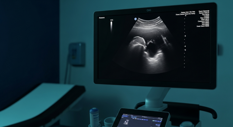
Imaging plays a pivotal role in detecting and characterising fatty liver disease. At IUS London, we utilise state-of-the-art ultrasound technology, often as the first-line investigation due to its non-invasive nature, accessibility, and ability to visualise liver parenchyma. The primary imaging modalities employed include:

Imaging ModalityDescriptionAdvantagesLimitations
**Ultrasound (US)**Initial imaging modality, detects moderate to severe steatosis as a bright, hyperechoic liver.Non-invasive, accessible, good for screening and identifying other pathologies.Less sensitive for mild steatosis, operator-dependent, cannot precisely quantify fat.
**Computed Tomography (CT)**Detects fatty liver as lower attenuation (darker) liver compared to the spleen.More sensitive than ultrasound for mild steatosis.Involves ionising radiation, not primary screening tool unless for other indications.
**Magnetic Resonance Imaging (MRI) with Proton Density Fat Fraction (PDFF)**Most accurate non-invasive method for quantifying liver fat, precisely measures fat percentage.Invaluable for diagnosis, monitoring progression, assessing treatment response, useful for equivocal ultrasound findings.Higher cost and accessibility compared to ultrasound.
**Transient Elastography (FibroScan)**Assesses liver stiffness, correlating with fibrosis; some devices offer Controlled Attenuation Parameter (CAP) for steatosis estimation.Used to stage NAFLD and identify risk of cirrhosis, complements other imaging.Does not directly measure fat, primarily assesses stiffness.

These imaging modalities, particularly advanced ultrasound and MRI, provide crucial insights into the presence and severity of fatty liver, guiding subsequent management strategies. At IUS London, our expert sonographers and radiologists are highly skilled in performing and interpreting these advanced scans, ensuring accurate and reliable diagnostic information.

Differential Diagnosis: Distinguishing Fatty Liver from Other Conditions

The diagnosis of fatty liver disease, particularly NAFLD, often requires careful consideration of other conditions that can present with similar symptoms, elevated liver enzymes, or imaging findings. A thorough differential diagnosis is crucial to ensure accurate patient management and to avoid misattributing symptoms to fatty liver when another primary liver pathology is present. At IUS London, our diagnostic process is designed to meticulously rule out these alternative or co-existing conditions.

Conditions Mimicking Fatty Liver on Imaging

While ultrasound, CT, and MRI are highly effective in detecting hepatic steatosis, certain patterns of fat deposition can mimic other liver pathologies, leading to diagnostic challenges:

  • Focal Fatty Sparing: This occurs when areas of the liver are spared from fat accumulation within a generally fatty liver. These spared areas can appear as hypodense (darker) lesions on CT or hyperechoic (brighter) lesions on ultrasound, potentially mimicking tumours or metastases. Advanced imaging, such as contrast-enhanced ultrasound or MRI, can help differentiate these benign findings from true lesions.
  • Focal Fatty Infiltration: Conversely, focal areas of fat accumulation within an otherwise normal liver can also appear as distinct lesions, sometimes mimicking neoplastic processes. Again, multiphasic imaging or MRI with fat suppression sequences are invaluable in confirming the fatty nature of these lesions.
  • Pseudotumours: In some cases, the pattern of fatty infiltration can be so atypical, forming nodular or mass-like lesions, that they are termed pseudotumours. These are particularly challenging and require careful correlation with clinical history and often further characterisation with advanced imaging to avoid unnecessary biopsies or interventions.

Other Liver Diseases with Similar Presentations

Several other liver conditions can cause elevated liver enzymes, fatigue, or abdominal discomfort, necessitating their exclusion before a definitive diagnosis of fatty liver disease is made. Viral Hepatitis (Hepatitis B and C) are chronic viral infections that are a common cause of liver inflammation and damage, often leading to elevated liver enzymes. Serological tests are essential to rule out these infections. Alcoholic Liver Disease (ALD), while distinct from NAFLD in its aetiology, can present with similar histological features such as steatosis, inflammation, and fibrosis. A detailed alcohol history is paramount in differentiating AFLD from NAFLD, though it is important to note that NAFLD and ALD can co-exist. Autoimmune Hepatitis is an inflammatory condition where the body's immune system attacks liver cells, causing significant liver enzyme elevations, and can be diagnosed through specific autoantibody tests and liver biopsy. Haemochromatosis is a genetic disorder leading to excessive iron absorption and deposition in the liver, causing damage, and is diagnosed by elevated iron studies and genetic testing. Wilson's Disease is a rare genetic disorder causing copper accumulation in the liver and other organs, diagnosed by low serum ceruloplasmin, elevated urinary copper, and genetic testing. Alpha-1 Antitrypsin Deficiency is a genetic condition that can lead to liver disease, particularly in adults, and is diagnosed by measuring alpha-1 antitrypsin levels. Drug-Induced Liver Injury (DILI) can be caused by many medications, both prescription and over-the-counter, as well as herbal supplements, making a comprehensive medication review essential. Coeliac Disease, while primarily an intestinal disorder, can sometimes manifest with elevated liver enzymes, which typically normalise with a gluten-free diet. Finally, Thyroid Disease, both hypothyroidism and hyperthyroidism, can cause mild elevations in liver enzymes.

Differential Diagnosis Table

To aid in understanding the distinctions, the following table summarises key differentiating features:

ConditionKey Differentiating FeaturesDiagnostic Approach
**NAFLD/NASH**Metabolic risk factors (obesity, diabetes, dyslipidaemia), minimal alcohol intake.Imaging (US, MRI-PDFF), blood tests (LFTs, glucose, lipids), exclusion of other causes.
**AFLD**History of significant alcohol consumption.Detailed alcohol history, often more severe liver enzyme elevations, imaging.
**Viral Hepatitis (B & C)**Specific viral markers in blood.Serological tests for HBsAg, anti-HCV.
**Autoimmune Hepatitis**Presence of autoantibodies (ANA, SMA, LKM-1), elevated IgG.Autoantibody panel, liver biopsy.
**Haemochromatosis**Elevated serum ferritin, transferrin saturation, genetic mutations (HFE).Iron studies, genetic testing.
**Wilson's Disease**Low serum ceruloplasmin, elevated urinary copper, Kayser-Fleischer rings.Copper studies, genetic testing.
**Drug-Induced Liver Injury**Temporal relationship with medication use, resolution upon drug withdrawal.Detailed medication history, drug cessation trial.

This comprehensive approach to differential diagnosis ensures that patients receive the most accurate diagnosis and appropriate management plan, tailored to their specific condition. At IUS London, our commitment to precision and patient well-being underpins every diagnostic step.

From Our Practice: A Clinical Insight at IUS London

[SUGGESTED — verify with clinician] A 48-year-old male, Mr. J.K., presented to IUS London for a routine health check-up, with no specific complaints. He had a history of well-controlled type 2 diabetes, managed with oral medication, and was moderately overweight (BMI 28). He reported occasional fatigue, which he attributed to his busy professional life. His liver function tests, performed by his GP, showed mildly elevated ALT (65 U/L) and AST (48 U/L), prompting a referral for an abdominal ultrasound. Our sonographer performed a detailed scan, which revealed diffuse hepatic steatosis, characterised by increased echogenicity of the liver parenchyma. Crucially, the ultrasound also identified a focal area of hypoechogenicity (appearing darker) in the right lobe of the liver, which raised initial concern for a potential mass. However, our experienced radiologist, correlating the findings with Mr. J.K.’s clinical history and the typical appearance of focal fatty sparing, recommended a follow-up MRI with PDFF. The MRI confirmed significant hepatic steatosis with a PDFF of 25% and definitively characterised the focal lesion as an area of focal fatty sparing, ruling out a tumour. This case highlights the importance of advanced imaging and expert interpretation in distinguishing benign variations of fatty liver from more serious pathologies, preventing unnecessary anxiety and invasive procedures. Mr. J.K. was advised on lifestyle modifications, including dietary changes and increased physical activity, to manage his fatty liver and diabetes, and was scheduled for a follow-up ultrasound in 6 months.

Book Your Private Ultrasound Scan at IUS London

If you are experiencing symptoms suggestive of liver issues, have risk factors for fatty liver disease, or simply wish to gain a comprehensive understanding of your liver health, IUS London offers state-of-the-art private ultrasound scans. Our team of highly skilled sonographers and radiologists provides accurate, timely, and compassionate diagnostic services in the heart of Kensington. Early detection and precise diagnosis are key to effective management and preventing the progression of liver disease. Take control of your health today.

Book your private ultrasound scan at IUS London

References

[1] Antunes, C. (2023). Fatty Liver - StatPearls. NCBI Bookshelf. Available at: https://www.ncbi.nlm.nih.gov/books/NBK441992/

[2] Vilgrain, V. (2013). Hepatic steatosis: A major trap in liver imaging. Diagnostic and Interventional Imaging, 94(10), 1029-1038. Available at: https://www.sciencedirect.com/science/article/pii/S2211568413000922

[3] Lăpădat, A. M., & Mărgăritescu, C. (2017). Non-invasive imaging techniques in assessing non-alcoholic fatty liver disease. Romanian Journal of Morphology and Embryology, 58(4), 1303-1308. Available at: https://pmc.ncbi.nlm.nih.gov/articles/PMC5304366/

[4] Wan, Y., et al. (2023). The imaging techniques and diagnostic performance of non-alcoholic fatty liver disease. Journal of Medical Imaging and Health Informatics, 13(3), 675-682. Available at: https://www.sciencedirect.com/science/article/pii/S168785072300136X

[5] Jang, W., et al. (2023). Non-Invasive Imaging Methods to Evaluate Non-Alcoholic Fatty Liver Disease. Diagnostics, 13(11), 1852. Available at: https://www.mdpi.com/2075-4418/13/11/1852

[6] Cleveland Clinic. (2023). Steatotic (Fatty) Liver Disease: Symptoms & Treatment. Available at: https://my.clevelandclinic.org/health/diseases/15831-fatty-liver-disease

[7] Mayo Clinic. Fatty liver disease (MASLD) - Symptoms and causes. Available at: https://www.mayoclinic.org/diseases-conditions/fatty-liver-disease-masld/symptoms-causes/syc-20354567

[8] British Liver Trust. Symptoms of liver disease. Available at: https://britishlivertrust.org.uk/information-and-support/liver-health-2/symptoms-of-liver-disease/

[9] Felix Hospital. When to See a Doctor for Fatty Liver: Know the Red Flags. Available at: https://www.felixhospital.com/blogs/when-to-see-a-doctor-for-fatty-liver-know-the-red-flags

[10] Mayo Clinic. Elevated liver enzymes: Causes. Available at: https://www.mayoclinic.org/symptoms/elevated-liver-enzymes/basics/causes/sym-20050830

[11] Cleveland Clinic. (2026). Elevated Liver Enzymes: Causes, Symptoms & Treatment. Available at: https://my.clevelandclinic.org/health/symptoms/17679-elevated-liver-enzymes

[12] Lee, T. H., & Kim, W. R. (2012). Evaluation of Elevated Liver Enzymes. Clinics in Liver Disease, 16(2), 237-246. Available at: https://pmc.ncbi.nlm.nih.gov/articles/PMC7110573/

[13] Wilkins, T., et al. (2013). Nonalcoholic Fatty Liver Disease: Diagnosis and Management. American Family Physician, 88(1), 35-42. Available at: https://www.aafp.org/afp/2013/0701/p35.html

[14] Yeh, M. M., & Brunt, E. M. (2007). Pathology of nonalcoholic fatty liver disease. American Journal of Clinical Pathology, 128(5), 837-846. Available at: https://academic.oup.com/ajcp/article-abstract/128/5/837/1760386

[15] Abd El-Kader, S. M., & El-Den Ashmawy, E. M. (2015). Non-alcoholic fatty liver disease: The diagnosis and management. World Journal of Hepatology, 7(6), 846-858. Available at: https://pmc.ncbi.nlm.nih.gov/articles/PMC4411527/

[16] Tannapfel, A., et al. (2011). Histopathological diagnosis of non-alcoholic and alcoholic fatty liver disease. Virchows Archiv, 459(5), 511-521. Available at: https://link.springer.com/article/10.1007/s00428-011-1066-1

Book Your Private Ultrasound Scan in London

Same-day appointments available in Kensington. No GP referral required. HCPC-registered consultant sonographers.