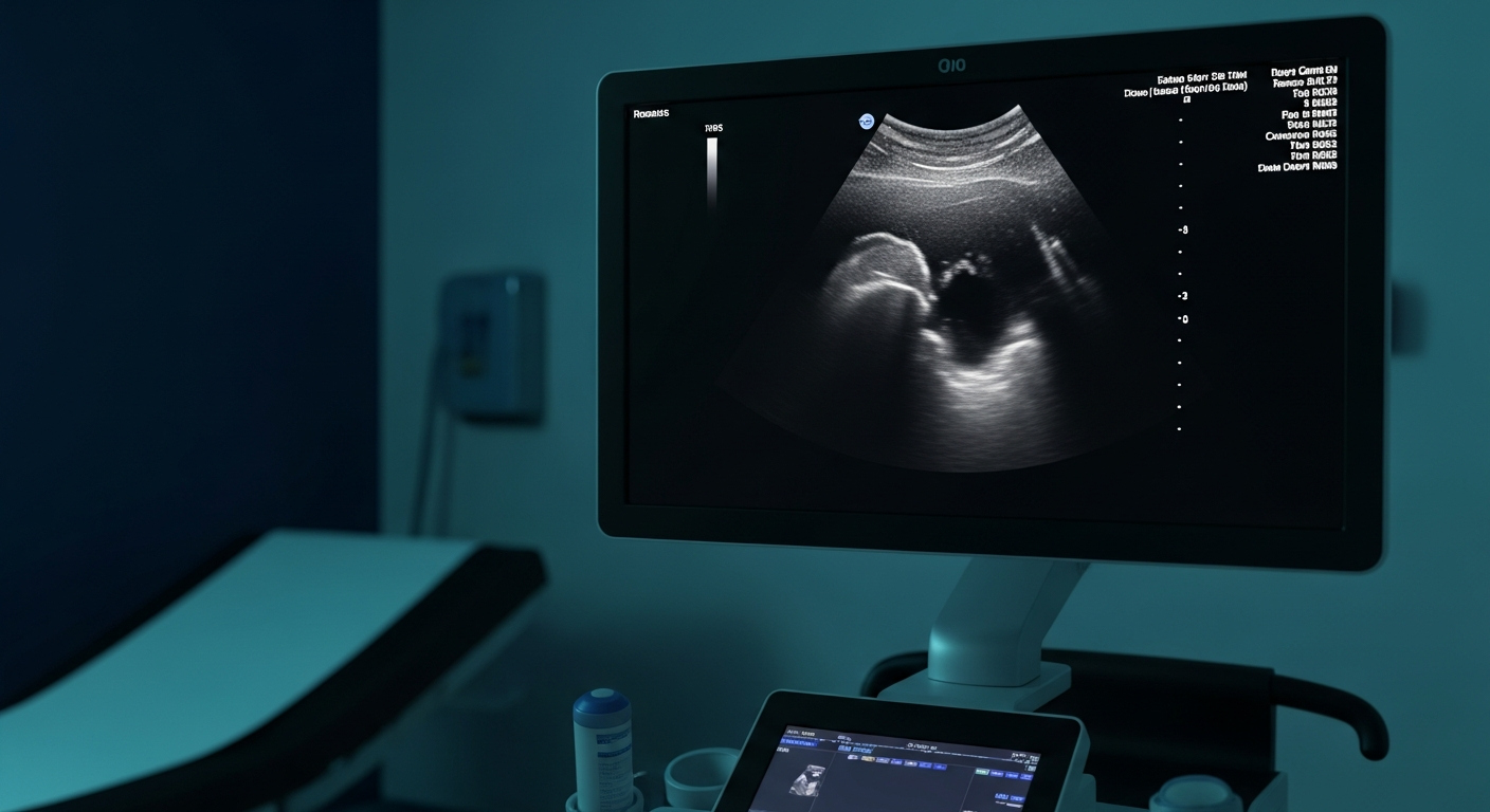
Liver Echotexture Explained — Heterogeneous, Coarse and Normal Findings

Liver Echotexture Explained: Heterogeneous, Coarse, and Normal

Unveiling the Liver's Story Through Sound: An Introduction to Echotexture

The liver, a vital organ nestled in the upper right quadrant of the abdomen, performs a myriad of essential functions, from detoxification and protein synthesis to bile production. Its health is paramount to overall well-being. When concerns about liver health arise, medical professionals often turn to non-invasive diagnostic tools, with ultrasound imaging standing out as a cornerstone. Ultrasound offers a window into the liver's internal architecture, providing crucial information about its size, shape, and, importantly, its echotexture.

Liver echotexture refers to the pattern of echoes reflected from the liver tissue as observed on an ultrasound scan. These echoes are generated when high-frequency sound waves, emitted by a transducer, encounter different tissue densities and structures within the liver. The way these sound waves are reflected, absorbed, or transmitted provides a visual representation that can indicate the presence of various liver conditions. A normal liver presents a characteristic, uniform appearance, while deviations from this norm can signal underlying pathology.

Understanding liver echotexture is not merely an academic exercise; it is a critical component of early detection, accurate diagnosis, and effective monitoring of liver health. Changes in echotexture can be the first indicators of conditions ranging from benign fatty liver disease to more serious issues like fibrosis, cirrhosis, or even focal lesions. For patients, comprehending what their echotexture report signifies can empower them to engage more actively in their healthcare journey and make informed decisions about their treatment and lifestyle.

This comprehensive article aims to demystify the complex terminology surrounding liver echotexture. We will delve into what constitutes a normal echotexture, explore the implications of increased echogenicity (often associated with fatty liver), and meticulously explain the significance of coarse and heterogeneous patterns. By the end of this discussion, both patients and healthcare enthusiasts will gain a clearer understanding of how ultrasound imaging helps tell the liver's story, guiding us towards better health outcomes.

The Fundamental Principles of Liver Ultrasound and Echotexture

To truly appreciate the nuances of liver echotexture, it is essential to grasp the basic principles behind ultrasound technology. Medical ultrasound operates on the principle of sonography, utilising high-frequency sound waves, typically in the range of 2 to 18 megahertz (MHz), to create real-time images of internal body structures. A handheld device called a transducer serves as both the emitter and receiver of these sound waves. When placed on the skin over the area of interest, the transducer sends sound waves into the body. These waves travel through tissues until they encounter an interface between different tissue types or structures. At these interfaces, some of the sound waves are reflected back to the transducer as echoes.

The strength and timing of these returning echoes are then processed by a computer to construct a two-dimensional image on a monitor. Tissues that reflect many sound waves appear bright on the screen (hyperechoic), while those that reflect fewer waves appear darker (hypoechoic) or black (anechoic, typically fluid-filled structures). The term echogenicity describes the brightness of tissues on an ultrasound image relative to surrounding structures or a reference organ.

Echotexture, specifically, refers to the overall pattern and uniformity of these echoes within a particular organ. For the liver, its echotexture is a visual representation of its internal cellular and structural composition. A healthy liver typically exhibits a fine, uniform, and homogeneous echotexture, meaning the echoes are evenly distributed and consistent throughout the organ. This uniform appearance is often compared to the texture of the renal cortex, providing a reliable internal reference point for radiologists and sonographers.

Several factors can influence the liver's echotexture, leading to variations from the normal homogeneous pattern. These include:

  • Tissue Density: Denser tissues tend to reflect more sound waves, appearing brighter.
  • Fat Content: Accumulation of fat within liver cells (steatosis) significantly increases echogenicity.
  • Fibrosis: The development of scar tissue (fibrosis) can alter the liver's architecture, leading to a coarser or more heterogeneous appearance.
  • Inflammation: Swelling and cellular infiltration due to inflammation can also impact echotexture.
  • Vascularity: The pattern of blood vessels within the liver contributes to its overall texture.

Understanding these influencing factors is crucial for interpreting ultrasound findings accurately. A change in echotexture is not a diagnosis in itself but rather an indicator that prompts further investigation and clinical correlation. It serves as a valuable clue, guiding healthcare providers toward identifying the underlying cause of liver abnormalities and formulating an appropriate management plan.

Normal Liver Echotexture: The Benchmark of Health

When a liver ultrasound report describes the echotexture as “normal,” it signifies a liver that is functioning optimally and appears healthy on imaging. A normal liver echotexture is characterized by a homogeneous, fine, mid-level echo pattern that is typically isoechoic (of similar brightness) or slightly hyperechoic compared to the renal cortex [8]. This means the internal structure of the liver appears uniform, without significant areas of increased brightness or darkness, and the echoes are finely dispersed.

Clinical Significance: A normal echotexture is a reassuring finding, indicating the absence of diffuse liver disease such as significant steatosis (fatty liver), widespread fibrosis, or cirrhosis. It suggests that the liver cells (hepatocytes) are healthy and the liver architecture is intact. For individuals undergoing routine check-ups or investigations for non-specific symptoms, a normal liver echotexture often helps rule out many common liver pathologies, thereby narrowing down diagnostic possibilities.

Patient Experience: Receiving a report stating “normal” liver echotexture can be a significant relief. It often means that any symptoms experienced are likely not related to a primary liver condition, or that an existing liver issue is well-managed and not causing structural changes detectable by ultrasound. This understanding can alleviate anxiety and provide a clear path forward for further investigations if symptoms persist, or simply reinforce the importance of maintaining a healthy lifestyle to preserve liver function.

Increased Echogenicity: The Hallmark of Fatty Liver Disease (Steatosis)

One of the most common abnormalities detected on liver ultrasound is increased echogenicity, often referred to as a “bright liver.” This finding is overwhelmingly associated with hepatic steatosis, more commonly known as fatty liver disease. Hepatic steatosis is characterized by the excessive accumulation of triglycerides (fat) within the hepatocytes, the primary cells of the liver. It is a global health concern, with its prevalence rising in parallel with the increasing rates of obesity, type 2 diabetes, and metabolic syndrome [3] [4] [6].

Clinical Explanation of Hepatic Steatosis

Definition and Pathophysiology: Hepatic steatosis occurs when the rate of fat accumulation in the liver exceeds the rate of fat metabolism and export. This can be due to various factors, including insulin resistance, excessive dietary intake of carbohydrates and fats, and certain medications. The presence of fat droplets within the liver cells alters the acoustic properties of the liver tissue. Fat is less dense than healthy liver parenchyma and scatters ultrasound waves more effectively, leading to a brighter appearance on the ultrasound image [5].

Grading of Steatosis: The severity of hepatic steatosis is typically graded based on the degree of increased echogenicity and other associated ultrasound features. While subjective, a common grading system includes:

  • Mild Steatosis: Slight, diffuse increase in fine echoes in the liver parenchyma with normal visualization of the diaphragm and intrahepatic vessel borders. The liver may appear slightly brighter than the renal cortex.
  • Moderate Steatosis: Moderate diffuse increase in fine echoes with slightly impaired visualization of the diaphragm and intrahepatic vessel borders. The liver is distinctly brighter than the renal cortex.
  • Severe Steatosis: Marked increase in fine echoes with poor or no visualization of the diaphragm and intrahepatic vessel borders, and often with posterior attenuation of the ultrasound beam. The liver appears very bright, almost white, on the scan [5] [8].

It is important to note that while ultrasound is highly sensitive for detecting moderate to severe steatosis, its sensitivity for mild steatosis can vary. Quantitative ultrasound techniques, which measure acoustic parameters like attenuation coefficient and backscatter coefficient, are emerging to provide more objective and precise quantification of hepatic steatosis [1] [6].

Patient Experience Walk-through with Fatty Liver Disease

For many individuals, hepatic steatosis is initially asymptomatic, meaning they experience no noticeable symptoms. The condition is often discovered incidentally during an ultrasound performed for other reasons, or as part of a routine health check-up. However, as the disease progresses, some patients may experience non-specific symptoms such as:

  • Fatigue: Persistent tiredness and lack of energy.
  • Dull ache or discomfort: A feeling of fullness or mild pain in the upper right abdomen.
  • Unexplained weight gain or difficulty losing weight.

Diagnosis: The diagnostic journey for fatty liver typically begins with an ultrasound. If increased echogenicity is noted, further investigations are usually recommended to confirm the diagnosis, assess the severity, and rule out other liver conditions. These may include blood tests to check liver enzymes (ALT, AST), lipid profiles, and markers for inflammation or fibrosis. In some cases, more advanced imaging techniques like MRI-PDFF (Magnetic Resonance Imaging Proton Density Fat Fraction) or liver biopsy may be performed to accurately quantify fat content and assess for inflammation and fibrosis, particularly if there is suspicion of non-alcoholic steatohepatitis (NASH), a more aggressive form of fatty liver disease [3].

Management: The cornerstone of managing hepatic steatosis, especially non-alcoholic fatty liver disease (NAFLD), involves lifestyle modifications. These include:

  • Dietary Changes: Adopting a balanced diet rich in fruits, vegetables, and whole grains, while limiting processed foods, sugary drinks, and unhealthy fats. A Mediterranean-style diet is often recommended.
  • Regular Exercise: Engaging in at least 150 minutes of moderate-intensity aerobic activity per week.
  • Weight Loss: Even a modest weight reduction (5-10% of body weight) can significantly improve liver fat and inflammation.
  • Alcohol Avoidance: For alcoholic fatty liver disease (AFLD), complete abstinence from alcohol is crucial.

Medical interventions may also be considered, particularly for patients with NASH or those at risk of progression to more severe liver disease. These can include medications to manage associated conditions like diabetes, high cholesterol, or hypertension.

Red Flags: When Increased Echogenicity Signals More Serious Concerns

While increased echogenicity often points to simple steatosis, it is crucial to be aware of red flags that might indicate a more serious underlying condition or progression of the disease. These include:

  • Persistent elevation of liver enzymes: While mild steatosis can cause slight elevations, significantly or persistently high liver enzymes warrant further investigation.
  • Signs of inflammation or fibrosis: If blood tests or other imaging (e.g., elastography) suggest inflammation or the development of scar tissue, it indicates progression to NASH or advanced liver disease.
  • Rapid onset or worsening of symptoms: Sudden fatigue, jaundice, or abdominal swelling are serious signs that require immediate medical attention.
  • Presence of other risk factors: Individuals with type 2 diabetes, severe obesity, or a family history of liver disease may be at higher risk for progression.

These red flags necessitate a thorough clinical review and potentially more invasive diagnostic procedures to accurately stage the disease and guide appropriate management. Early recognition and intervention are key to preventing the progression to cirrhosis and its complications.

Coarse Liver Echotexture: A Sign of Chronic Change

Beyond simple increased echogenicity, another significant finding on liver ultrasound is a coarse liver echotexture. This term describes an irregular, non-uniform pattern of echoes within the liver parenchyma, often indicative of chronic and diffuse changes in the liver architecture. Unlike the fine, homogeneous pattern of a healthy liver or the uniformly bright appearance of fatty liver, a coarse echotexture suggests a more disrupted internal structure [2] [9] [10].

Clinical Explanation of Coarse Echotexture

Definition and Underlying Causes: A coarse echotexture typically arises from widespread alterations in the liver tissue, most commonly due to fibrosis and cirrhosis. Fibrosis is the excessive accumulation of extracellular matrix proteins, primarily collagen, in response to chronic liver injury. If the injury persists, fibrosis can progress to cirrhosis, a severe and irreversible form of liver scarring characterized by widespread nodules and distortion of the liver architecture. These structural changes disrupt the normal passage of ultrasound waves, leading to the irregular, coarse appearance.

Common underlying causes of coarse liver echotexture include:

  • Chronic Viral Hepatitis: Long-term infections with Hepatitis B (HBV) or Hepatitis C (HCV) viruses are leading causes of chronic liver inflammation, fibrosis, and cirrhosis worldwide.
  • Alcohol-Related Liver Disease (ARLD): Chronic excessive alcohol consumption can lead to a spectrum of liver damage, from fatty liver to alcoholic hepatitis and cirrhosis.
  • Non-Alcoholic Steatohepatitis (NASH): As mentioned earlier, NASH is an advanced form of NAFLD where fat accumulation is accompanied by inflammation and liver cell damage, which can lead to fibrosis and cirrhosis.
  • Autoimmune Liver Diseases: Conditions like autoimmune hepatitis, primary biliary cholangitis (PBC), and primary sclerosing cholangitis (PSC) cause chronic inflammation and scarring.
  • Metabolic and Genetic Disorders: Hemochromatosis (iron overload), Wilson's disease (copper overload) [2], and alpha-1 antitrypsin deficiency can also lead to chronic liver damage and fibrosis.

Progression: The development of coarse echotexture often reflects a progressive disease process. It signifies that the liver has been subjected to prolonged injury, leading to attempts at repair that result in scar tissue formation. This progression can be conceptualized as a continuum:

1. Inflammation: Initial injury leads to inflammation.

2. Fibrosis: Persistent inflammation triggers the deposition of collagen and other extracellular matrix components, forming scar tissue.

3. Cirrhosis: Extensive fibrosis leads to the formation of regenerative nodules and severe distortion of the liver architecture, impairing liver function.

Patient Experience Walk-through with Coarse Echotexture

Patients with coarse liver echotexture may experience a range of symptoms, depending on the underlying cause and the stage of liver disease. In early stages of fibrosis, symptoms may be subtle or non-existent. However, as fibrosis progresses to cirrhosis, more pronounced symptoms can emerge, including:

  • Fatigue and Weakness: Profound and persistent tiredness.
  • Jaundice: Yellowing of the skin and eyes due to impaired bilirubin processing.
  • Ascites: Accumulation of fluid in the abdomen, causing swelling.
  • Peripheral Edema: Swelling in the legs and ankles.
  • Easy Bruising or Bleeding: Due to reduced production of clotting factors.
  • Hepatic Encephalopathy: Confusion, disorientation, and memory problems due to the liver's inability to remove toxins from the blood.
  • Portal Hypertension: Increased pressure in the portal vein, leading to complications like varices (enlarged veins) in the esophagus and stomach.

Diagnosis: When a coarse echotexture is identified on ultrasound, further diagnostic steps are crucial to determine the extent of liver damage and the underlying cause. These may include:

  • Blood Tests: Comprehensive liver function tests, viral hepatitis serology, autoimmune markers, and tests for genetic conditions.
  • Elastography: Techniques like Transient Elastography (TE, FibroScan) or Sound Touch Viscoelastography (STVi) [2] measure liver stiffness, which correlates with the degree of fibrosis. These non-invasive methods are increasingly used to stage fibrosis and monitor disease progression.
  • Advanced Imaging: CT or MRI scans may be used to provide more detailed anatomical information, assess for complications of cirrhosis, and rule out other pathologies.
  • Liver Biopsy: Historically the gold standard, liver biopsy involves taking a small tissue sample for microscopic examination to assess the degree of inflammation, fibrosis, and the specific cause of liver disease. While less frequently performed due to advances in non-invasive methods, it remains valuable in certain cases.

Management: Management of coarse echotexture and the underlying chronic liver disease focuses on addressing the root cause and managing complications. This can involve:

  • Antiviral Therapy: For chronic viral hepatitis.
  • Abstinence from Alcohol: For ARLD.
  • Immunosuppressants: For autoimmune liver diseases.
  • Dietary and Lifestyle Modifications: To support liver health and manage symptoms.
  • Management of Complications: Diuretics for ascites, beta-blockers for portal hypertension, and medications for hepatic encephalopathy.
  • Liver Transplant: In cases of end-stage liver disease.

Red Flags: Signs of Decompensated Liver Disease

For patients with coarse echotexture, particularly those with known chronic liver disease, certain red flags indicate the progression to decompensated cirrhosis, a life-threatening stage requiring urgent medical attention:

  • New onset or worsening jaundice.
  • Development or worsening of ascites.
  • Gastrointestinal bleeding (e.g., vomiting blood or black, tarry stools) from esophageal varices.
  • Confusion, disorientation, or changes in mental status (hepatic encephalopathy).
  • Recurrent infections.
  • Kidney dysfunction (hepatorenal syndrome).

These symptoms signify a critical decline in liver function and necessitate immediate medical evaluation and intervention.

Heterogeneous Liver Echotexture: Context is Key

The term heterogeneous liver echotexture describes a liver that appears patchy, uneven, or inconsistent in its echo pattern. This means there are varying areas of brightness and darkness, or different textures, interspersed throughout the liver parenchyma. Unlike the diffuse uniformity seen in normal or even fatty livers, heterogeneity suggests a more localized or irregular distribution of changes within the liver. The interpretation of heterogeneous echotexture is highly context-dependent, requiring careful correlation with the patient's clinical history, symptoms, and other diagnostic findings.

Clinical Explanation of Heterogeneous Echotexture

Definition and Causes: A heterogeneous echotexture can arise from a multitude of conditions, ranging from benign findings to more serious pathologies. The key characteristic is the lack of uniform echo distribution. Common causes include:

* Cysts: Fluid-filled sacs, typically anechoic (black) with posterior enhancement.

* Hemangiomas: Benign vascular tumors, often hyperechoic (bright).

* Adenomas or Focal Nodular Hyperplasia (FNH): Benign solid tumors.

* Malignant Tumors: Primary liver cancers (hepatocellular carcinoma) or metastatic lesions, which can be hypoechoic, hyperechoic, or mixed.

  • Focal Lesions: This is a frequent cause of heterogeneity. The liver may contain discrete areas that differ in echogenicity from the surrounding parenchyma. These can include:
  • Acute Inflammation: In some cases of acute hepatitis, the liver may appear diffusely heterogeneous due to edema and inflammatory infiltrates.
  • Localized Fatty Infiltration or Sparing: Fat deposition in the liver is not always uniform. Some areas may be heavily infiltrated with fat, while others are spared, leading to a patchy, heterogeneous appearance. Conversely, areas of normal liver tissue within a diffusely fatty liver can appear hypoechoic (darker) relative to the surrounding bright, fatty liver, a phenomenon known as focal fat sparing.
  • Advanced Diffuse Liver Disease: While coarse echotexture is more typical of diffuse fibrosis/cirrhosis, in some advanced cases, particularly with regenerative nodules or areas of necrosis, the liver can also appear heterogeneous.
  • Post-Procedural Changes: Changes following liver biopsy, surgery, or interventional procedures can also result in localized areas of altered echotexture.

Importance of Clinical Correlation: Given the wide range of potential causes, a finding of heterogeneous echotexture on its own is rarely diagnostic. It serves as a prompt for further investigation. The sonographer or radiologist will look for additional features, such as the presence of a mass, its borders, internal vascularity, and any associated changes in liver size or contour. This information, combined with the patient's medical history (e.g., history of cancer, viral hepatitis, alcohol use), symptoms, and blood test results, is crucial for accurate interpretation.

Patient Experience Walk-through with Heterogeneous Echotexture

Receiving an ultrasound report that mentions “heterogeneous liver echotexture” can be a source of significant anxiety and uncertainty for patients. The ambiguity of the term often leads to concerns about serious conditions, particularly cancer. It is vital for healthcare providers to communicate clearly that heterogeneity is a descriptive term, not a diagnosis, and that further investigation is almost always warranted to determine its cause.

Anxiety and Uncertainty: Patients may experience a range of emotions, from mild concern to significant distress, especially if they have a family history of liver disease or cancer. The waiting period for further tests can be particularly stressful.

Further Investigations: Depending on the suspected cause, additional imaging modalities are typically employed to characterize the heterogeneous findings. These may include:

  • Contrast-Enhanced Ultrasound (CEUS): Can provide more detailed information about blood flow patterns within focal lesions.
  • Computed Tomography (CT) Scan: Offers cross-sectional images that can better delineate lesions and assess their relationship to surrounding structures.
  • Magnetic Resonance Imaging (MRI) with Liver-Specific Contrast: Considered the gold standard for characterizing many liver lesions, providing excellent soft tissue contrast and functional information.
  • Liver Biopsy: In cases where imaging is inconclusive or malignancy is suspected, a biopsy may be performed to obtain tissue for histological examination.

Follow-up: For benign findings, such as small hemangiomas or focal fat sparing, regular follow-up ultrasounds may be recommended to monitor for any changes. For more significant findings, a clear management plan will be developed based on the definitive diagnosis.

Red Flags: Suspicious Focal Lesions and Rapid Changes

While many causes of heterogeneous echotexture are benign, certain red flags necessitate prompt and thorough evaluation:

  • Suspicious Focal Lesions: Any new or growing mass, especially if it has irregular borders, internal vascularity, or atypical enhancement patterns on advanced imaging, raises concern for malignancy.
  • Rapid Changes in Liver Function Tests: A sudden and unexplained deterioration in liver enzyme levels or synthetic function (e.g., albumin, INR) in conjunction with heterogeneous echotexture warrants urgent investigation.
  • Constitutional Symptoms: Unexplained weight loss, persistent fever, night sweats, or severe fatigue accompanying heterogeneous echotexture can be indicative of a more serious underlying disease process, such as malignancy or infection.
  • History of Cancer: In patients with a known history of cancer, a new heterogeneous finding in the liver must be thoroughly evaluated for metastatic disease.

These red flags underscore the importance of a multidisciplinary approach, involving radiologists, hepatologists, oncologists, and surgeons, to ensure accurate diagnosis and timely management.

⚠ Red Flags: When to Seek Expert Opinion

Understanding liver echotexture is a critical step in assessing liver health, but it is equally important to know when to seek expert medical advice. While this article provides a comprehensive overview, it is not a substitute for professional medical consultation. Any abnormal finding on a liver ultrasound, whether it's increased echogenicity, coarse, or heterogeneous echotexture, warrants a thorough evaluation by a qualified healthcare professional.

Summary of Key Indicators for Concern:

  • Persistent or Worsening Symptoms: Fatigue, unexplained weight loss, abdominal pain or swelling, jaundice, dark urine, or pale stools.
  • Abnormal Liver Function Tests: Elevated liver enzymes (ALT, AST, GGT, ALP), bilirubin, or abnormal clotting factors.
  • New or Changing Ultrasound Findings: Any new mass, significant increase in echogenicity, or progression of coarse/heterogeneous patterns.
  • Known Risk Factors: History of chronic viral hepatitis, significant alcohol consumption, obesity, diabetes, metabolic syndrome, or family history of liver disease.

Importance of Regular Check-ups and Follow-up: For individuals with known liver conditions or risk factors, regular medical check-ups and adherence to recommended follow-up imaging schedules are paramount. This allows for early detection of changes, timely intervention, and monitoring of disease progression. Proactive management can significantly impact long-term outcomes and prevent the development of severe complications.

The Role of Specialist Consultation: When an abnormal liver echotexture is identified, consultation with a specialist, such as a hepatologist (a liver specialist) or a gastroenterologist, is often recommended. These experts possess in-depth knowledge of liver diseases and can provide a definitive diagnosis, develop a personalized treatment plan, and coordinate further investigations. They can also offer guidance on lifestyle modifications, medication management, and, if necessary, discuss advanced treatment options, including liver transplantation.

The Patient Journey: From Ultrasound to Understanding

Navigating a potential liver health concern can be daunting. Understanding the typical patient journey, from the initial ultrasound to comprehending the results and managing a diagnosis, can help alleviate anxiety and empower individuals to be active participants in their care.

What to Expect During a Liver Ultrasound

  • Preparation: You will typically be asked to fast for 6-8 hours before the scan. This helps to reduce gas in the abdomen, which can interfere with image quality, and ensures the gallbladder is distended for better visualization. You may also be asked to wear loose, comfortable clothing.
  • Procedure: The ultrasound is a non-invasive and painless procedure. You will lie on an examination table, and a clear gel will be applied to your abdomen. This gel helps the transducer make good contact with your skin and allows sound waves to travel effectively. The sonographer will then move the transducer across your abdomen, capturing images of your liver from various angles. You may be asked to hold your breath briefly or change positions to optimize image acquisition. The scan usually takes 15-30 minutes.

Interpreting Your Results: How to Understand the Report

Once the ultrasound is complete, a radiologist will interpret the images and generate a report. This report will describe the findings, including the liver's size, shape, presence of any focal lesions, and, of course, its echotexture. It is crucial to discuss this report with your referring doctor or specialist. They will explain the medical terminology in plain language, correlate the findings with your symptoms and other test results, and outline the next steps.

Key aspects to understand in your report:

  • Echotexture Description: Pay attention to terms like “homogeneous,” “hyperechoic,” “coarse,” or “heterogeneous.”
  • Focal Lesions: If any masses or abnormalities are noted, the report will describe their characteristics and suggest further imaging if needed.
  • Associated Findings: The report may also comment on other organs, such as the gallbladder, bile ducts, or spleen, which can provide additional context.

Coping with a Diagnosis: Emotional Support and Resources

Receiving a diagnosis related to liver health can be emotionally challenging. It is important to remember that you are not alone and resources are available to help you cope:

  • Open Communication with Your Doctor: Do not hesitate to ask questions and express your concerns. A clear understanding of your condition is the first step towards effective management.
  • Support Groups: Connecting with others who have similar experiences can provide emotional support, practical advice, and a sense of community.
  • Lifestyle Changes: Focusing on positive lifestyle changes, such as diet and exercise, can give you a sense of control and actively contribute to your liver health.
  • Mental Health Support: If you are struggling with anxiety, depression, or stress related to your diagnosis, consider seeking support from a mental health professional.

From Our Practice: IUS London’s Approach to Liver Health

At IUS London, we understand that a liver ultrasound is often more than just an imaging procedure; it's a crucial step in a patient's health journey. Our commitment extends beyond providing high-quality diagnostic images to offering a holistic, patient-centric approach to liver health. We pride ourselves on combining cutting-edge technology with the expertise of highly skilled specialists, ensuring that every patient receives the most accurate diagnosis and a clear path forward.

Our Expertise: Advanced Ultrasound Techniques and Experienced Specialists

Our practice is equipped with the latest ultrasound technology, allowing for detailed visualization and characterization of liver echotexture and any associated pathologies. Our sonographers and radiologists are not only proficient in standard B-mode ultrasound but are also experienced in advanced techniques such as:

  • Elastography (e.g., Shear Wave Elastography): This non-invasive technique measures liver stiffness, providing a quantitative assessment of fibrosis, which is crucial for staging chronic liver disease and monitoring treatment response. It offers a valuable alternative or adjunct to liver biopsy.
  • Contrast-Enhanced Ultrasound (CEUS): By using microbubble contrast agents, CEUS enhances the visualization of blood flow within liver lesions, aiding in the differentiation between benign and malignant masses without the need for ionizing radiation or nephrotoxic contrast agents.

Our team comprises specialists with extensive experience in hepatology and diagnostic imaging, ensuring that complex cases are interpreted with precision and clinical insight. We are dedicated to continuous learning and staying abreast of the latest advancements in liver imaging and disease management.

Patient-Centric Care: Personalized Treatment Plans and Holistic Approach

We believe that effective healthcare is deeply personal. Every patient's liver health journey is unique, and our approach reflects this philosophy. When you come to IUS London, you can expect:

  • Thorough Consultation: A detailed discussion of your medical history, symptoms, and concerns before any procedure.
  • Clear Communication: We ensure that all findings are explained in an understandable manner, empowering you with the knowledge to make informed decisions.
  • Personalized Management Plans: Based on your ultrasound findings, clinical context, and individual needs, we collaborate with you and your referring physician to develop a tailored management strategy. This may involve further investigations, lifestyle recommendations, or referral to other specialists.
  • Holistic Support: We recognize that liver health is intertwined with overall well-being. Our approach considers not just the physical aspects of your condition but also the emotional and psychological impact, offering support and resources to help you navigate your health journey.

Case Studies: Real-World Impact at IUS London

To illustrate our commitment to patient care and the practical application of our expertise, consider these anonymized examples from our practice:

  • Case Study 1: Early Detection of Steatosis: Mrs. S, a 45-year-old with newly diagnosed type 2 diabetes, underwent a routine liver ultrasound at IUS London. The scan revealed mild increased echogenicity, indicative of early hepatic steatosis. Through personalized dietary and exercise recommendations, and close monitoring, Mrs. S was able to reverse her fatty liver, preventing progression to more severe disease.
  • Case Study 2: Characterizing a Heterogeneous Finding: Mr. J, a 60-year-old with a history of chronic hepatitis C, presented with a heterogeneous liver echotexture on a screening ultrasound. Our advanced CEUS and subsequent MRI confirmed a small hepatocellular carcinoma, allowing for early intervention and successful treatment, significantly improving his prognosis.
  • Case Study 3: Monitoring Chronic Liver Disease: Ms. A, a 55-year-old with known non-alcoholic steatohepatitis (NASH), regularly attends IUS London for elastography. This non-invasive monitoring has allowed us to track the progression of her liver fibrosis accurately, guiding her treatment adjustments and providing reassurance about the stability of her condition.

These examples highlight how our comprehensive approach, from precise diagnostics to personalized care, makes a tangible difference in the lives of our patients.

Comprehensive FAQs about Liver Echotexture

To further clarify common questions and concerns regarding liver echotexture, we have compiled a list of frequently asked questions:

What is the difference between coarse and heterogeneous echotexture?

While both terms describe variations from a normal homogeneous liver, they refer to distinct patterns. Coarse echotexture typically implies a diffuse, irregular, and somewhat granular appearance throughout the liver, often associated with widespread fibrosis or cirrhosis due to chronic liver disease. It suggests a global alteration of the liver's internal architecture. In contrast, heterogeneous echotexture describes a patchy or uneven appearance, where different areas of the liver have varying echo patterns. This can be due to localized issues like focal lesions (cysts, tumors), areas of focal fat infiltration or sparing, or acute inflammation. The key distinction lies in the distribution: coarse is generally diffuse, while heterogeneous can be more localized or patchy.

Can liver echotexture change over time?

Yes, liver echotexture can absolutely change over time, and these changes often reflect the dynamic nature of liver health. For instance, increased echogenicity due to fatty liver (steatosis) can often be reversed or significantly improved with lifestyle modifications such as weight loss, dietary changes, and increased physical activity. Similarly, in some cases of early fibrosis, if the underlying cause of liver injury is removed or effectively treated, there can be a regression of fibrosis, which might be reflected in an improvement in echotexture. However, advanced fibrosis and cirrhosis, which typically manifest as coarse echotexture, are often irreversible, though progression can be halted or slowed with appropriate management. Regular follow-up ultrasounds are crucial for monitoring these changes.

Is an abnormal echotexture always serious?

Not necessarily. An abnormal echotexture is an indicator that warrants further investigation, but it does not automatically mean a serious condition. For example, mild increased echogenicity due to fatty liver is very common and, in many cases, can be managed effectively with lifestyle changes without leading to severe complications. Similarly, some causes of heterogeneous echotexture, such as small benign cysts or focal fat sparing, are not serious. However, other abnormal findings, particularly coarse echotexture or suspicious focal lesions within a heterogeneous liver, can indicate significant liver disease or potentially serious conditions. The seriousness depends on the specific pattern, the presence of other symptoms, blood test results, and the patient's overall clinical picture. It is always best to consult with a healthcare professional for an accurate assessment.

What lifestyle changes can improve liver echotexture?

For conditions like fatty liver disease (increased echogenicity), significant improvements in echotexture can be achieved through lifestyle modifications:

  • Healthy Diet: Emphasize whole foods, fruits, vegetables, lean proteins, and healthy fats. Limit processed foods, sugary drinks, and excessive saturated/trans fats. A Mediterranean-style diet is often recommended.
  • Regular Exercise: Aim for at least 150 minutes of moderate-intensity aerobic activity or 75 minutes of vigorous-intensity activity per week, combined with strength training.
  • Weight Management: Losing even a modest amount of weight (5-10% of body weight) can significantly reduce liver fat.
  • Limit Alcohol: Reduce or eliminate alcohol consumption, especially if you have fatty liver or other liver conditions.
  • Manage Underlying Conditions: Effectively manage conditions like diabetes, high cholesterol, and hypertension, as they can contribute to liver disease.

These changes can reduce fat accumulation, decrease inflammation, and potentially reverse early fibrosis, leading to an improvement in liver echotexture.

How often should I have a liver ultrasound if I have abnormal echotexture?

The frequency of follow-up ultrasounds depends on the specific diagnosis, the severity of the condition, and your overall clinical picture. For mild, stable fatty liver, your doctor might recommend an ultrasound every 1-2 years, or as needed based on symptoms or blood test results. If there is evidence of more advanced disease (e.g., moderate to severe steatosis, fibrosis, or cirrhosis), or if you have suspicious focal lesions, more frequent monitoring (e.g., every 6-12 months) may be necessary. Your specialist will determine the most appropriate follow-up schedule based on established clinical guidelines and your individual risk factors.

What other tests might be needed after an abnormal ultrasound?

An abnormal liver ultrasound often serves as a starting point for further investigation. Depending on the findings, your doctor may recommend:

  • Blood Tests: Comprehensive liver function tests, viral hepatitis serology, autoimmune markers, iron studies, and genetic tests.
  • Elastography: Non-invasive tests like FibroScan to assess liver stiffness and quantify fibrosis.
  • Advanced Imaging: CT scans or MRI with liver-specific contrast to further characterize focal lesions or assess the extent of diffuse disease.
  • Liver Biopsy: In select cases, a liver biopsy may be performed to obtain a tissue sample for microscopic examination, especially when the diagnosis is unclear or precise staging of fibrosis and inflammation is required.

These additional tests help to establish a definitive diagnosis, assess disease severity, and guide treatment decisions.

Conclusion: Empowering You with Knowledge for Liver Health

The liver, a silent workhorse of the human body, communicates its health status through various signals, and liver echotexture on ultrasound is one of the most insightful. From the reassuring homogeneity of a normal liver to the bright echoes of steatosis, the coarse patterns of chronic change, and the varied landscape of heterogeneity, each finding tells a part of the liver's story. Understanding these nuances empowers individuals to engage proactively with their health, recognize potential red flags, and seek timely medical attention.

At IUS London, we are dedicated to providing clarity and comprehensive care in the realm of liver diagnostics. Our advanced ultrasound capabilities, combined with the expertise of our specialists, ensure that you receive not just an image, but a thorough understanding of your liver health. We believe that informed patients are empowered patients, capable of making the best decisions for their well-being.

Take Control of Your Liver Health Today

Concerned about your liver health? Do you have questions about a recent ultrasound report, or are you experiencing symptoms that might indicate a liver issue? Don't leave your health to chance. Contact IUS London today for an expert ultrasound assessment and a personalized care plan. Our team is here to guide you through every step of your journey towards optimal liver health. Visit our website to learn more about our services and schedule your consultation.

References

1. Nam, K., et al. (2023). Improving diagnostic accuracy of ultrasound texture features in detecting and quantifying hepatic steatosis using various beamforming sound speeds. Physics in medicine and biology. https://consensus.app/papers/details/ded101aa850b5ba885b8b69151397a8a/

2. Li, Y., et al. (2025). Assessment of liver involvement in Wilson’s disease with different liver echo patterns based on liver stiffness evaluated on Transient elastography and Sound Touch Viscoelastography. Scientific Reports. https://consensus.app/papers/details/cb292ba5f2055db7b97cca8f45348130/

3. Pirmoazen, A. M., et al. (2020). Quantitative ultrasound approaches for diagnosis and monitoring hepatic steatosis in nonalcoholic fatty liver disease. Theranostics. https://consensus.app/papers/details/8155990ab93a58c491211bef0e0003aa/

4. Khov, N., et al. (2014). Bedside ultrasound in the diagnosis of nonalcoholic fatty liver disease. World journal of gastroenterology. https://consensus.app/papers/details/85a437d599385e92904419b2b697f4b1/

5. Palmentieri, B., et al. (2006). The role of bright liver echo pattern on ultrasound B-mode examination in the diagnosis of liver steatosis. Digestive and liver disease : official journal of the Italian Society of Gastroenterology and the Italian Association for the Study of the Liver. https://consensus.app/papers/details/80955e7c1a035c1fb18d4e7eca22af3a/

6. Park, J., et al. (2022). Quantitative Evaluation of Hepatic Steatosis Using Advanced Imaging Techniques: Focusing on New Quantitative Ultrasound Techniques. Korean Journal of Radiology. https://consensus.app/papers/details/4f5ddab8bb7759f3a1253234f5d0d4c6/

7. Baek, J., et al. (2021). Clusters of ultrasound scattering parameters for the classification of steatotic and normal livers. Ultrasound in medicine & biology. https://consensus.app/papers/details/c7acee9d118f53ecb6cc59e190edf103/

8. Tan, Z., et al. (2024). Hepatic steatosis: Qualitative and quantitative sonographic assessment in comparison to histology. Australasian Journal of Ultrasound in Medicine. https://consensus.app/papers/details/a05397dc6f125fafb379471604b50b65/

9. Destrempes, F., et al. (2022). Quantitative ultrasound, elastography, and machine learning for assessment of steatosis, inflammation, and fibrosis in chronic liver disease. PLoS ONE. https://consensus.app/papers/details/f0a062736fbb575f85fd9783786e2be2/

10. Mathiesen, U., et al. (2002). Increased liver echogenicity at ultrasound examination reflects degree of steatosis but not of fibrosis in asymptomatic patients with mild/moderate abnormalities of liver transaminases. Digestive and liver disease : official journal of the Italian Society of Gastroenterology and the Italian Association for the Study of the Liver. https://consensus.app/papers/details/48f68c0a47ff576b9080109819179090/

Internal Links & Call to Action

  • Explore Our Services: Discover how IUS London can support your liver health journey. Visit our Liver Ultrasound Services page.
  • Learn More About Fatty Liver Disease: Get in-depth information on diagnosis, management, and prevention. Read our guide on Fatty Liver Disease.
  • Ready to take the next step? Contact IUS London today to schedule your expert ultrasound assessment and receive a personalized care plan. Your liver health is our priority. priority.

Book Your Private Ultrasound Scan in London

Same-day appointments available in Kensington. No GP referral required. HCPC-registered consultant sonographers.