Monitoring Fibroids — How Often Should I Be Scanned?
Uterine fibroids are a common, non-cancerous growth that develops in the uterus. While many women with fibroids experience no symptoms, others may suffer from heavy menstrual bleeding, pelvic pain, pressure, or fertility issues. The decision to monitor fibroids and the frequency of scans depend on several factors, including the size and location of the fibroids, the presence and severity of symptoms, and the patient's age and reproductive plans.
Understanding Uterine Fibroids: Anatomy, Pathology, and Growth Mechanisms
Uterine fibroids, medically termed leiomyomas or myomas, are the most common benign tumours of the female reproductive tract, originating from the smooth muscle cells (myocytes) of the uterine wall [1]. These growths are characterised by the proliferation of uterine smooth muscle cells embedded within an abundant extracellular matrix [2]. Their size can range from microscopic seedlings to large masses that significantly distort the uterus, sometimes filling the entire pelvic cavity [3].
Fibroids are categorised based on their anatomical location within the uterus, which often dictates the type and severity of symptoms experienced:
- Intramural fibroids: These are the most common type, developing within the muscular wall (myometrium) of the uterus. They can enlarge the uterus and lead to symptoms such as heavy bleeding and pelvic pressure.
- Subserosal fibroids: Located on the outer surface of the uterus, these fibroids can grow outwards, potentially pressing on adjacent organs like the bladder or bowel, causing pressure symptoms. They can also be pedunculated, meaning they are attached to the uterus by a stalk.
- Submucosal fibroids: These fibroids protrude into the uterine cavity (endometrial cavity). Even small submucosal fibroids can cause significant symptoms, including heavy and prolonged menstrual bleeding (menorrhagia) and infertility, as they can interfere with embryo implantation [3] [4]. They can also be pedunculated.
- Cervical fibroids: Less common, these develop in the wall of the cervix.
Pathogenesis and Growth
The precise aetiology of uterine fibroids is complex and multifactorial, involving a delicate interplay of genetic, hormonal, and environmental influences [5]. Key drivers of fibroid growth include:
- Hormonal Influence: Oestrogen and progesterone are critical in promoting fibroid growth. These steroid hormones stimulate the proliferation of smooth muscle cells and the deposition of extracellular matrix within the fibroid [6]. Fibroids possess a higher concentration of oestrogen and progesterone receptors compared to normal myometrial tissue, making them highly responsive to hormonal fluctuations throughout the menstrual cycle. This explains why fibroids typically grow during the reproductive years and often regress or shrink after menopause when these hormone levels naturally decline [1].
- Genetic Predisposition: A significant proportion of fibroids exhibit chromosomal abnormalities and genetic mutations. For instance, mutations in the *MED12* gene are frequently identified in fibroid tissue, playing a crucial role in their development and growth [7]. Other genetic factors, such as overexpression of *HMGA2* and fumarate hydratase deficiency, also contribute to fibroid pathobiology [8].
- Growth Factors and Extracellular Matrix: Fibroid growth is not solely dependent on cellular proliferation but also on the excessive accumulation of extracellular matrix components, such as collagen and fibronectin [2] [6]. Various growth factors, including transforming growth factor-beta (TGF-β) and insulin-like growth factor (IGF), are implicated in this process, fostering a microenvironment conducive to fibroid expansion [9].
- Epigenetic Mechanisms: Emerging research highlights the role of epigenetic modifications, such as DNA methylation and microRNAs, in regulating gene expression within fibroid cells, further contributing to their abnormal growth patterns [10].
Understanding these underlying mechanisms is crucial for developing targeted therapies and improving management strategies for uterine fibroids.
Why Monitor Fibroids?
Monitoring fibroids is crucial for several reasons:
1. Symptom Management: Tracking changes in fibroid size and location can help correlate with symptom severity and guide treatment decisions.
2. Growth Assessment: While most fibroids grow slowly, rapid growth can sometimes indicate a rare cancerous change (leiomyosarcoma), though this is exceedingly uncommon.
3. Fertility and Pregnancy: Fibroids can impact fertility and pregnancy outcomes. Monitoring is essential for women planning conception or those who are pregnant.
4. Treatment Planning: Regular monitoring helps determine the optimal timing and type of intervention if treatment becomes necessary.
How Often Should I Be Scanned?
The frequency of scans for monitoring fibroids is highly individualised. For asymptomatic fibroids, where there are no bothersome symptoms, a common recommendation is to monitor every 6-12 months. This typically involves a transvaginal ultrasound, which provides clear images of the uterus and fibroids.
However, the monitoring schedule should be adjusted if:
- New symptoms develop: The onset or worsening of symptoms such as heavy bleeding, severe pain, or pressure warrants an earlier re-scan.
- Rapid growth is suspected: While rare, a significant increase in fibroid size may necessitate further investigation.
- Pregnancy is planned or occurs: Close monitoring is often required during pregnancy due to hormonal changes that can influence fibroid growth.
Diagnostic Tools for Fibroid Monitoring: A Patient's Perspective
Monitoring uterine fibroids often involves various imaging techniques, each offering unique insights into the fibroids' size, location, and characteristics. Understanding what to expect during each type of scan can help alleviate anxiety and ensure a smoother experience.
Transvaginal Ultrasound (TVS)
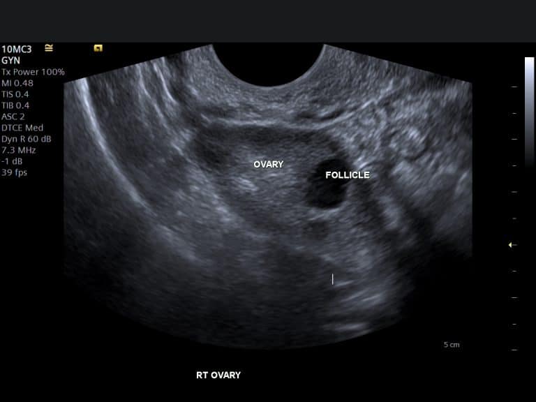
What it is: The transvaginal ultrasound is the most common and often the first-line imaging modality for evaluating uterine fibroids. It uses high-frequency sound waves emitted from a small probe inserted into the vagina to create detailed images of the uterus, ovaries, and surrounding pelvic structures. This proximity allows for excellent resolution and precise measurements of fibroids, especially those located within the uterine wall or protruding into the cavity.
The Patient Experience:
1. Preparation: Typically, no special preparation is required. You may be asked to empty your bladder before the scan to improve image clarity.
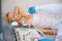
2. Procedure: You will lie on an examination table, similar to a gynaecological exam. A thin, lubricated probe, slightly larger than a tampon, will be gently inserted into your vagina. The sonographer will move the probe to visualise different parts of your uterus and ovaries. You might feel some pressure, but it should not be painful. If you experience discomfort, please inform the sonographer immediately.
3. Duration: The scan usually takes between 15 to 30 minutes.
4. Benefits: It is a quick, non-invasive (in terms of internal penetration, but not surgical), and cost-effective method. It provides real-time images, allowing for immediate assessment of fibroid size, number, and location.
Abdominal Ultrasound
What it is: An abdominal ultrasound uses sound waves transmitted through a transducer placed on the abdomen to generate images of the pelvic organs. This method is particularly useful for larger fibroids that may extend beyond the pelvis and are difficult to fully visualise with a transvaginal approach.
The Patient Experience:
1. Preparation: You will typically be asked to drink several glasses of water before the scan to ensure a full bladder. A full bladder helps push the bowel loops out of the way, providing a clearer window to view the uterus and fibroids.
2. Procedure: You will lie on your back on an examination table. A clear gel will be applied to your lower abdomen, and the sonographer will move a transducer (a small, handheld device) across your skin. You might feel some pressure as the transducer is moved, especially with a full bladder.
3. Duration: This scan usually lasts about 20 to 40 minutes.
4. Benefits: It is completely non-invasive and can provide a broader view of the pelvic area, making it suitable for very large fibroids or when a transvaginal scan is not feasible or comfortable.
Magnetic Resonance Imaging (MRI)
What it is: MRI is a sophisticated imaging technique that uses a powerful magnetic field and radio waves to create highly detailed cross-sectional images of soft tissues, including the uterus and fibroids. It offers superior soft tissue contrast compared to ultrasound and is often used for surgical planning, when ultrasound findings are inconclusive, or to differentiate fibroids from other uterine masses.
The Patient Experience:
1. Preparation: You may be asked to avoid food and drink for a few hours before the scan. You will need to remove all metallic objects, including jewellery, watches, and any clothing with metal fasteners, as these can interfere with the magnetic field. Inform the radiographer if you have any metallic implants (e.g., pacemakers, certain IUDs, surgical clips).
2. Procedure: You will lie on a movable table that slides into a large, tunnel-like machine. It is crucial to remain still during the scan. You will hear loud knocking or buzzing noises, so earplugs or headphones will be provided. In some cases, a contrast agent may be injected intravenously to enhance the visibility of certain structures.
3. Duration: An MRI scan can take anywhere from 30 to 60 minutes, or sometimes longer, depending on the complexity of the imaging required.
4. Benefits: MRI provides the most comprehensive anatomical detail of fibroids, including their exact size, number, and precise location relative to other pelvic organs. It can also help in characterising fibroid tissue, which is vital for treatment planning, especially for minimally invasive procedures like uterine fibroid embolisation.
From Our Practice
At IUS London, we believe in a personalised approach to fibroid management, recognising that each woman's experience with fibroids is unique. Our strategy focuses on watchful waiting, regular monitoring, and tailored interventions when necessary. Here are a few anonymised clinical scenarios from our practice that illustrate our approach:
Scenario 1: Watchful Waiting with Stable Asymptomatic Fibroids
- Patient Profile: Mrs. A, a 45-year-old professional, was incidentally found to have multiple small intramural fibroids (largest 3 cm) during a routine gynaecological check-up. She reported no symptoms such as heavy bleeding, pain, or pressure.
- Our Approach: Given her asymptomatic status and the relatively small size of the fibroids, we recommended a watchful waiting approach with annual transvaginal ultrasounds. Over five years, her fibroids have remained stable in size, and she has continued to be symptom-free. This consistent monitoring has allowed her to avoid invasive treatments, providing peace of mind and maintaining her quality of life.
Scenario 2: Symptomatic Fibroids Requiring Closer Monitoring and Intervention Discussion
- Patient Profile: Ms. B, a 32-year-old woman, presented with increasingly heavy and prolonged menstrual bleeding, leading to significant fatigue and iron-deficiency anaemia. Ultrasound revealed a 6 cm submucosal fibroid.
- Our Approach: Due to her debilitating symptoms and the location of the fibroid, which is known to cause heavy bleeding, we initiated a more frequent monitoring schedule (every 6 months) to track fibroid growth and assess the impact of conservative management strategies (e.g., hormonal therapy to reduce bleeding). We also engaged in detailed discussions about potential interventions, including hysteroscopic myomectomy (a minimally invasive procedure to remove submucosal fibroids) once her anaemia was managed. Our aim was to alleviate her symptoms while preserving her fertility potential.
Scenario 3: Fibroids in the Context of Fertility Planning
- Patient Profile: Mrs. C, a 38-year-old hoping to conceive, had a history of a 4 cm intramural fibroid located near the fallopian tube. She had experienced difficulty conceiving for over a year.
- Our Approach: We conducted a detailed MRI scan to precisely map the fibroid's location and its potential impact on her uterine cavity and fallopian tubes. Given her fertility concerns, we discussed the option of laparoscopic myomectomy to remove the fibroid, which could improve her chances of natural conception. Post-surgery, we continued to monitor her uterine healing and fibroid recurrence with regular ultrasounds as she pursued her fertility journey.
These cases highlight the importance of individualised care, where monitoring frequency and treatment decisions are carefully tailored to each patient's unique clinical picture, symptoms, and life goals.
Frequently Asked Questions (FAQs) About Fibroid Monitoring
Here are some common questions and concerns patients often have regarding uterine fibroids and their monitoring:
Q1: Are fibroids always symptomatic?
A: No, a significant number of women with uterine fibroids experience no symptoms at all. These are often discovered incidentally during routine pelvic examinations or imaging for other conditions. The presence and severity of symptoms depend largely on the fibroid's size, number, and, most importantly, its location within the uterus. For instance, submucosal fibroids, even if small, are more likely to cause heavy bleeding due to their proximity to the uterine lining, whereas intramural or subserosal fibroids might need to grow quite large before causing noticeable symptoms like pressure or pain.
Q2: Can fibroids turn cancerous?
A: It is extremely rare for uterine fibroids to become cancerous. Fibroids are benign (non-cancerous) growths. In very rare cases, a cancerous tumour called a leiomyosarcoma can develop in the uterus. These are distinct from fibroids and do not typically arise from pre-existing fibroids. Rapid growth of a fibroid, especially after menopause, can sometimes raise suspicion and warrant further investigation, but this is an uncommon occurrence.
Q3: What factors influence fibroid growth?
A: Fibroid growth is primarily influenced by hormones, particularly oestrogen and progesterone. These hormones stimulate fibroid cells to grow and multiply. This is why fibroids tend to grow during the reproductive years and often shrink after menopause when hormone levels decline. Other factors include genetic predisposition, certain growth factors, and extracellular matrix components that contribute to their development and expansion.
Q4: Is there anything I can do to prevent fibroids from growing or developing?
A: While there's no guaranteed way to prevent fibroids, some lifestyle factors may play a role. Maintaining a healthy weight, eating a balanced diet rich in fruits and vegetables, and regular exercise may help. Some studies suggest that vitamin D deficiency might be linked to fibroid development, so ensuring adequate vitamin D levels could be beneficial. However, more research is needed in this area.
Q5: What are the alternatives to surgery for fibroid management?
A: For many women, especially those with mild or no symptoms, watchful waiting with regular monitoring is a primary management strategy. Medical treatments, such as hormonal therapies (e.g., birth control pills, GnRH agonists), can help manage symptoms like heavy bleeding. Non-surgical procedures like Uterine Fibroid Embolisation (UFE) can shrink fibroids by blocking their blood supply. The best approach depends on your symptoms, fibroid characteristics, and future reproductive plans.
Q6: How do fibroids affect pregnancy and fertility?
A: Fibroids can impact fertility and pregnancy outcomes, though many women with fibroids have successful pregnancies. Submucosal fibroids, which protrude into the uterine cavity, are most likely to interfere with embryo implantation and increase the risk of miscarriage. During pregnancy, fibroids can sometimes grow due to hormonal changes, potentially leading to pain, premature labour, or complications during delivery. Close monitoring is essential for pregnant women with fibroids.
Q7: What should I do if I experience new or worsening symptoms?
A: If you experience any new or worsening symptoms, such as unusually heavy or prolonged bleeding, severe pelvic pain, increased pressure, or any other concerning changes, it is crucial to contact your doctor or specialist immediately. These symptoms may indicate a change in your fibroids or another underlying condition that requires prompt medical attention. Your monitoring schedule may need to be adjusted, and further investigations might be necessary.
⚠ Red Flags
While fibroids are generally benign, certain signs warrant immediate medical attention:
- Sudden, severe pelvic pain.
- Unexplained heavy or prolonged vaginal bleeding.
- Rapid increase in fibroid size, especially after menopause.
- Symptoms of anaemia (fatigue, shortness of breath, dizziness) due to heavy bleeding.
References
[1] American College of Obstetricians and Gynecologists. (2021). Uterine Fibroids. Retrieved from https://www.acog.org/womens-health/faqs/uterine-fibroids
[2] Dolmans, M. M. (2024). Pathogenesis of uterine fibroids: current understanding. Fertility and Sterility, 121(4), 627-628. https://www.sciencedirect.com/science/article/pii/S0015028224001699
[3] Barjon, K. (2025). Uterine Leiomyomata. StatPearls. https://www.ncbi.nlm.nih.gov/books/NBK546680/
[4] Navarro, A. (2021). Understanding the Impact of Uterine Fibroids on Human Endometrium. International Journal of Molecular Sciences, 22(11), 5865. https://pmc.ncbi.nlm.nih.gov/articles/PMC8186666/
[5] UpToDate. (2025). Uterine fibroids (leiomyomas): Histology and pathogenesis. https://www.uptodate.com/contents/uterine-fibroids-leiomyomas-histology-and-pathogenesis/print
[6] Yang, Q. (2022). Comprehensive Review of Uterine Fibroids. Endocrine Reviews, 43(4), 678-709. https://academic.oup.com/edrv/article/43/4/678/6422392
[7] Bulun, S. E. (2025). Uterine fibroids. Physiological Reviews, 105(1), 1-35. https://journals.physiology.org/doi/abs/10.1152/physrev.00010.2024
[8] Ramaiyer, M. S. (2024). Genetic Mechanisms Driving Uterine Leiomyoma Pathobiology, Epidemiology, and Treatment. Genes, 15(5), 558. https://www.mdpi.com/2073-4425/15/5/558
[9] Munro, M. G. (2025). The epidemiology and pathogenesis of uterine fibroids. International Journal of Gynecology & Obstetrics, 150(2), 155-162. https://obgyn.onlinelibrary.wiley.com/doi/full/10.1002/ijgo.70527
[10] Baranov, V. S. (2019). Pathogenomics of uterine fibroids development. International Journal of Molecular Sciences, 20(24), 6151. https://www.mdpi.com/1422-0067/20/24/6151
Call to Action
If you are experiencing symptoms related to uterine fibroids or have concerns about your fibroid monitoring schedule, contact IUS London today to schedule a consultation with our specialists. We are dedicated to providing comprehensive and compassionate care tailored to your individual needs.
Book Your Private Ultrasound Scan in London
Same-day appointments available in Kensington. No GP referral required. HCPC-registered consultant sonographers.