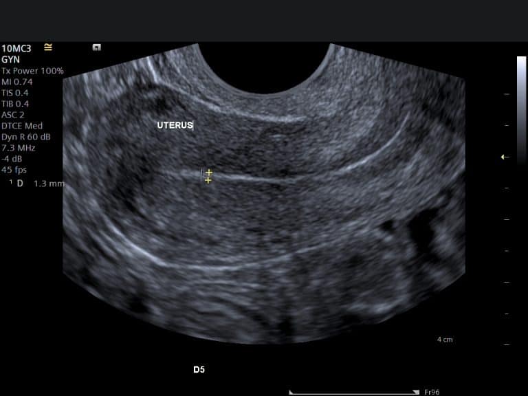
Pelvic Pain — When Is It Time for a Scan?
Pelvic pain is one of the most common reasons women seek help — and one of the most often dismissed. Here is what the pattern of pain suggests, which situations need same-day care, and what an ultrasound scan can and cannot find.

Some pelvic pain is a normal part of a cycle. Some is a structural problem that a scan finds in twenty minutes. And a small amount needs a hospital today, not an appointment next week. The useful skill is telling those apart — which mostly comes down to how the pain arrived, whether it tracks your cycle, and what comes with it.
When pelvic pain needs same-day care
Sudden severe one-sided pain, pain with a positive pregnancy test, pain with fever, or pain with fainting or collapse needs assessment today — call NHS 111, contact your GP urgently, or go to A&E. These situations belong with the NHS emergency pathway, not a booked scan.
What the pattern of pain suggests
Pain that tracks your cycle. A sharp twinge mid-cycle is often ovulation itself and settles within a day or two. Pain concentrated around periods that is getting worse over time — especially with pain during sex or with bowel movements at period time — is the classic pattern of endometriosis or adenomyosis, and deserves proper assessment rather than stronger painkillers.
Pain that ignores your cycle. A constant ache or pressure, bloating that does not come and go, or pain on one side that persists points more towards ovarian cysts, fibroids or — less often — a previous infection having left its mark. Persistent bloating with pelvic pain lasting more than a few weeks is specifically worth a GP visit; our guide to pelvic ultrasound when CA-125 is raised explains how that assessment fits together.
Pain from somewhere else. The bladder, bowel and the muscles and joints of the pelvis all refer pain to the same place. Pain tied to urination, to eating, or to movement and posture is real pelvic pain — it is just less likely to have a gynaecological cause, which changes who should look at it first.
What a pelvic ultrasound can — and cannot — find
| Ultrasound sees well | Ultrasound cannot see |
|---|---|
| Ovarian cysts — and whether they look simple, complex or like an endometrioma | Superficial endometriosis — a normal scan does not exclude it |
| Fibroids: how many, how large, and where they sit | Adhesions (internal scarring) in most cases |
| Features of adenomyosis in the womb muscle | Bowel causes such as IBS |
| A dilated fallopian tube, free fluid, and the womb lining | Bladder pain syndrome, muscle, joint and nerve pain |
That cuts both ways. A scan that finds a cause gives you something concrete to act on. A normal scan meaningfully narrows the list — and it does not mean the pain is imagined. It means the cause is likely one of the things ultrasound cannot show, and the next step is a different kind of assessment, not no assessment.
Scan first, or GP first?
A pelvic scan is a sensible first step when the pain is recurring or cyclical, when you are watching a known cyst or fibroids, or when period pain has changed character and you want the structural questions answered before deciding what to do next. You do not need a referral to book.
Your GP is the right first stop when bladder or bowel symptoms dominate, when there is unexplained weight loss or fever, or when pain comes with bleeding between periods or after sex — those combinations need an examination and sometimes tests a scan cannot replace. And any of the same-day situations at the top of this page go straight to the urgent pathway.
What the scan involves
- 1
BookingNo referral needed. We confirm preparation when you book — it differs depending on whether the scan is transabdominal or transvaginal.
- 2
The scanA transvaginal scan gives the clearest view of the womb and ovaries and is the standard approach; a transabdominal scan is available if you prefer. The appointment is 30 minutes.
- 3
What we assessThe womb and its lining, both ovaries, the tissues around them, and any cyst or fibroid measured and described properly — including whether it explains your pain.
- 4
ResultsFindings are explained on screen as we scan, and the written report is usually with you within two hours — ready for your GP or gynaecologist if follow-up is needed.
Your questions answered
Can ultrasound see endometriosis?
My scan was normal but I am still in pain. What now?
Do I need a full bladder for the scan?
Is pain in the middle of my cycle normal?
When is pelvic pain an emergency?
Can the scan find bowel-related causes?
Where to go from here
What counts as heavy, what causes it, and what a scan can find.
Read the guide →
FibroidsMonitoring FibroidsWhen fibroids need watching and how surveillance works.
Read the guide →
TestsRaised CA-125Why a pelvic ultrasound belongs alongside the blood test.
Read the guide →
Looked at properly, explained clearly
A full pelvic assessment — womb, lining and both ovaries — with every finding measured, explained on screen at the appointment, and the written report usually with you within two hours.
Kensington, London W8 4ED
3 mins from Notting Hill Gate
This article is general information about pelvic pain, not individual medical advice. Sudden severe pain, pain with a positive pregnancy test, fever, or fainting needs same-day NHS care. For persistent pain, see your GP whatever a scan shows — NHS guidance on pelvic pain sets out the causes and pathway.
Written and clinically reviewed by the HCPC-registered sonographers who perform this scan at IUS London — a CQC-registered diagnostic ultrasound clinic (Provider ID 1-2775844974). Your own findings are explained to you at the scan and set out in your report.
Author: Yianni Kiromitis, Senior Sonographer, HCPC RA38415 — over 20 years’ experience in NHS and private ultrasound
Medically reviewed: 9 August 2026