UTIs in Women — and What an Ultrasound Can Actually Show
A scan cannot diagnose a urinary infection. What it can do is find the reason they keep coming back — which is a different and often more useful question.
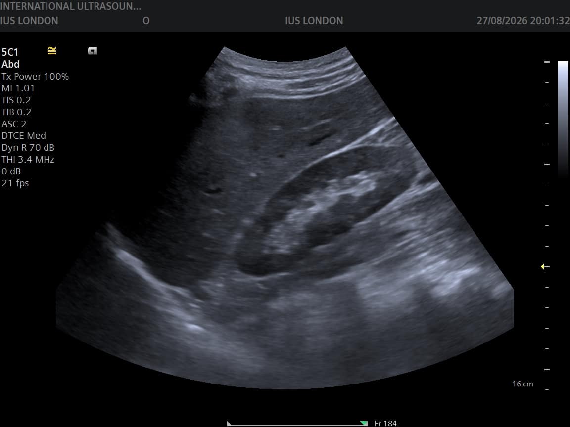
Let us answer the question most people arrive with, immediately and plainly: no, an ultrasound cannot detect a urinary tract infection. Infection is diagnosed from your urine and your symptoms — a dipstick test, and where needed a laboratory culture that identifies the bacteria and which antibiotics will work. Ultrasound cannot see bacteria, and inflamed bladder lining often looks entirely normal on a scan. If you have burning, urgency and frequency, the useful appointment is with your GP or pharmacist, not with us.
So why does this page exist? Because there is a second question underneath the first, and it is the one ultrasound answers well: why do these infections keep happening? For recurrent or complicated UTIs, imaging looks for a structural reason — and finding one changes the treatment from another round of antibiotics into something that actually resolves the pattern.
A temperature of 38°C or above, shivering or chills, pain in your back or side beneath the ribs, nausea or vomiting, blood in your urine, or new confusion — particularly in an older person. These suggest the infection has reached the kidneys (pyelonephritis) or is becoming systemic, and need same-day assessment by your GP, NHS 111 or A&E. Do not book a scan and wait.
What actually diagnoses a UTI
| Test | What it tells you |
|---|---|
| Symptoms | Burning on passing urine, urgency, frequency, lower abdominal discomfort, cloudy or strong-smelling urine. In otherwise healthy women with classic symptoms, this alone often justifies treatment |
| Urine dipstick | Detects nitrites and white blood cells suggesting infection — quick, done in the surgery |
| Urine culture | Identifies which bacterium is responsible and which antibiotics it responds to. Important for recurrent infections, in pregnancy, or when treatment has failed |
| Ultrasound | Does not diagnose the infection. Looks for structural causes and complications behind recurrent or complicated cases |
Women are more prone to UTIs for anatomical reasons — a shorter urethra, positioned closer to the bowel — and Escherichia coli from the bowel is the usual culprit. Sexual activity, menopause and the drop in oestrogen, diabetes, and anything that stops the bladder emptying fully all increase the likelihood.
What ultrasound looks for in recurrent UTIs
Recurrent UTI generally means two or more infections in six months, or three or more in a year. At that point the question shifts from “is there an infection?” to “why does this keep happening?” — and a urinary tract scan examines the kidneys, the bladder, and how well the bladder empties, looking for:
- Incomplete bladder emptying — measured as the post-void residual, the volume left behind after you pass urine. Residual urine is a reservoir for bacteria and one of the commonest findings that explains a recurrent pattern
- Kidney or bladder stones — which harbour bacteria and resist antibiotics until dealt with
- Obstruction and hydronephrosis — swelling of the kidney when urine cannot drain freely
- Structural variation — a duplex system, a bladder diverticulum, or other anatomy that predisposes to infection
- Complications of infection — scarring, or a collection of pus requiring urgent treatment
- Bladder wall thickening — a non-specific sign that may reflect chronic irritation
A normal scan is genuinely useful information too: it makes an anatomical cause unlikely and points management towards prevention — fluid intake, post-menopausal vaginal oestrogen, and where appropriate a preventive antibiotic strategy, all discussed with your GP.
When a scan is worth having — and when it is not
Worth having: recurrent infections meeting the pattern above; infection that has not responded to appropriate antibiotics; visible blood in the urine; suspected stones; a kidney infection that is not settling; or persistent urinary symptoms with repeatedly negative urine tests, where something other than infection may be responsible.
Not worth having: a single straightforward UTI with typical symptoms and a good response to treatment. Scanning that adds nothing, and we would rather say so than take the booking. Equally, do not use a scan to decide whether you have an infection right now — it cannot tell you, and the delay matters.
Timing tip: if you are being scanned for recurrent UTI, it is generally more informative once the acute infection has been treated, so the findings reflect your underlying anatomy rather than temporary inflammation. Your GP can advise, and can also refer you on the NHS — worth asking about before booking privately.
What the scan involves
A urinary tract scan takes about 30 minutes and is performed through the abdomen — no internal examination. You arrive with a comfortably full bladder so the bladder can be assessed when full; you are then asked to empty it and rescanned briefly, which is how the post-void residual is measured. That before-and-after pair is the part that most often explains a recurrent pattern, and it is why the appointment is arranged the way it is.
Findings are explained on screen as they are found, and a written report follows — usually within two hours — phrased so your GP can act on it directly. Where something needs onward referral, the report says so plainly.
Your questions answered
Can an ultrasound detect a UTI?
Can you see a bladder infection on a scan?
Why has my GP requested an ultrasound for recurrent UTIs?
What is a post-void residual?
How many UTIs count as recurrent?
Should I have the scan while I have an infection?
What if my urine tests keep coming back negative but symptoms persist?
Where to go from here
Finding out why they keep coming back
Kidneys, bladder and post-void residual assessed by HCPC-registered sonographers, explained on screen, with a written report usually within two hours for your GP. For an infection you have right now, see your GP — not us.
Kensington, London W8 4ED
3 mins from Notting Hill Gate
This article is general information, not individual medical advice. Suspected urinary tract infection should be assessed by your GP or pharmacist, and symptoms suggesting a kidney infection need same-day medical assessment. Imaging investigates the causes of recurrent infection; it does not diagnose infection itself.
Written and clinically reviewed by the HCPC-registered sonographers who perform this scan at IUS London — a CQC-registered diagnostic ultrasound clinic (Provider ID 1-2775844974). Your own findings are explained to you at the scan and set out in your report.
Author: Yianni Kiromitis, Senior Sonographer, HCPC RA38415
Medically reviewed: 1 August 2026