Table of Contents
It is quite common for our London pregnant couples to have their first pregnancy scan at around 7 weeks of pregnancy. The most common reasons for this private ultrasound scan is to confirm the pregnancy, to check viability and that everything is OK in general.
Other reasons to have a 7-week baby ultrasound are to:
- Confirm whether it is a singleton or multiple pregnancies.
- Confirm gestational age
- Spotting or bleeding
- Confirm the presence of a heartbeat.
- Measure the foetal pole and ensure the size of the baby is the right size for gestational age.
- Make sure the uterus, fallopian tubes and ovaries are OK.
- Confirm the foetal pole is within the endometrial cavity and it is not an ectopic pregnancy
- When a mother has irregular periods and is therefore unsure about her gestational age.
When is a dating scan necessary?
This is a scan or ultrasound which determines your expected date of delivery (EDD) based on the development of the embryo.
A dating scan is generally done for women who:
- Are unsure about their last menstrual period.
- Have irregular cycles
- Recently had a miscarriage and have soon conceived again.
- Stopped using hormonal contraception.
- Have conceived while they are breastfeeding.
- In any other situation such as multiple partners, where confirming gestational age is considered important.
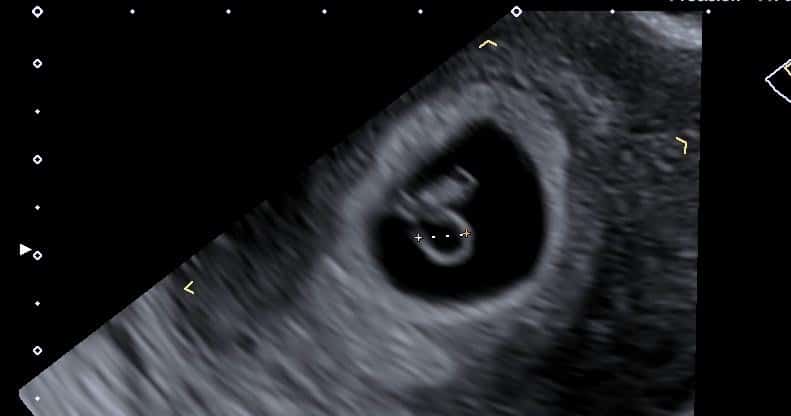
How big will my baby be at the 7-week ultrasound?
The average crown to rump length (CRL) of the embryo at 7 weeks gestation is between 5mm-12mm.
Crown/rump length and gestational age are closely compared with each other during pregnancy ultrasound until around the end of the first trimester.
When should I have my earliest scan?
The ideal time to assess the gestation age with ultrasound is between 7-10 weeks of pregnancy. The crown-rump length measurements obtained at this gestational age can vary between 3-5 days.
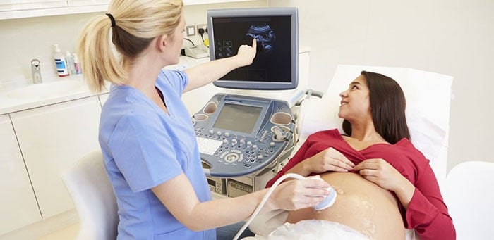
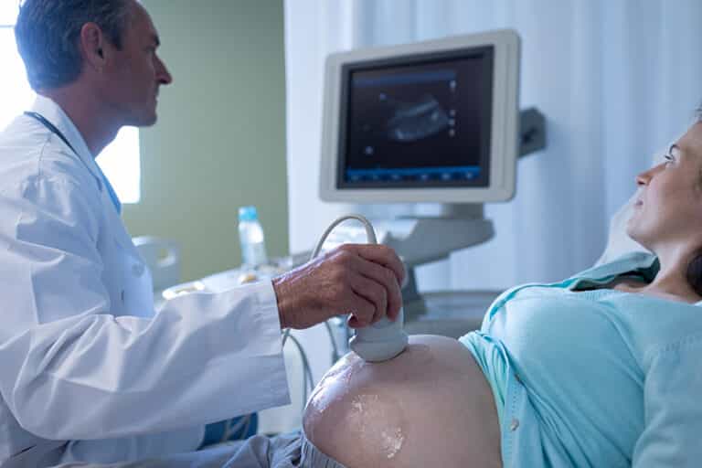
How will my 7-week ultrasound be done?
This early baby scan is normally performed trans-abdominally. In some instances, however, an internal scan (Transvaginal) may be required to see all the necessary detail or if your womb tilts backwards.
We will always try to scan trans-abdominally first but if we need to do a transvaginal ultrasound scan then we will discuss this with you. To perform this early baby scan, you will be asked to lie down on the examination couch and expose your lower abdomen.
A small amount of water-based gel will be applied to your skin. The gel will help the transducer to make good contact with the skin. The ultrasound transducer will be placed on the body and will be moved in different directions over the area of interest to obtain the required information/ultrasound images.
There is usually no discomfort from pressure as the transducer or probe as otherwise known, is pressed against the area being examined. However, if scanning is performed over an area of tenderness, you may feel pressure or minor discomfort from the transducer.
Once the ultrasound scan is completed, the clear ultrasound gel will be wiped off your skin. Any portions that are not wiped off will dry quickly. The ultrasound gel does not usually stain or discolour clothing.
This ultrasound examination is usually completed within 10-15 minutes. After an ultrasound examination, you should be able to resume your normal activities immediately.
Can I see my baby's heartbeat at the seven-week ultrasound?
Heartbeat should be visible at 6+ weeks gestation and you should be able to see a tubular flickering structure. The baby's heart rate at this early stage of pregnancy is fast and it is between 90-110 beats per minute.
Will my seven week ultrasound be really clear?
The image quality of your pregnancy ultrasound scan will depend on the quality of the ultrasound equipment and also the experience and skills of the sonographer.
At International Ultrasound Services we have years of ultrasound scanning experience and therefore you can rest assured that we will achieve the best image quality possible without compromising the safety of the baby or mother.
If you have any questions please leave a comment and we will do our best to answer.
At our private ultrasound clinic, we offer pregnancy scans from as early as 5-6 weeks in times to suit you.
Who interprets the results of the early pregnancy scan and how do I get them?
Our Sonographer, a Health Care Professional specifically trained to perform and understand the ultrasound images, will do your exam and provide you with a written report that you can take it to your doctor. Our sonographers will also discuss the results with you during and after your examination.
It is very common that sometimes when you leave you remember of a question that you forgot to ask, so please don't hesitate to contact us either via phone or e-mail and we will do our best to answer your question.
What are the benefits and risks of the early pregnancy ultrasound examination?
Benefits
- The early pregnancy, reassurance scan is non-invasive.
- If a transvaginal scan is required the ultrasound exam might be a little uncomfortable but not painful.
- Ultrasound examinations are significantly cheaper than other diagnostic imaging modalities.
- Ultrasound imaging is safe for the baby and the mother as there is no ionizing radiation involved.
- You can not see the baby using conventional x-ray imaging.
- Ultrasound is the preferred imaging investigation for the diagnosis and monitoring of pregnancies.
- Ultrasound allows the sonographer to see inside the uterus in real-time and provides necessary information about the pregnancy.
Risks
- There are no harmful effects on humans or babies related to pregnancy ultrasound examinations.
- Although ultrasound has been used in pregnancy for more than 40 years with no evidence suggesting it is harmful to the patient, embryo or fetus, ultrasound should be performed only when clinically indicated and by qualified practitioners.
About Pregnancy Scans
A pregnancy ultrasound scan is the same as a ‘normal' scans but is being used to evaluate the overall health of your baby instead of looking at other organs such as gallbladder for gallstones or kidney for kidney stones. So in pregnancy ultrasound scans are being used to visualise the baby, the placenta, the uterus and cervix and your ovaries.
Pregnancy ultrasound scans or prenatal ultrasounds are very common and being carried in any stage of the pregnancy.
When Is an Ultrasound Performed During Pregnancy in the NHS?
You will normally be offered two ultrasound scans during your pregnancy from the NHS. The first pregnancy scan is at 12 weeks and called a dating scan. The second pregnancy scan is at 20 weeks and called an anomaly scan.
Most of the expectant mothers, especially the ones with previous complications such as miscarriage they do not believe that 2 ultrasound scans during pregnancy are enough and this is the reason they choose to have a private pregnancy scan in London with us.
What are ultrasound scans used for in pregnancy?
Depending on your stage of pregnancy, ultrasounds will be used to give you and your doctor or midwife answers about your pregnancy.
First Trimester Ultrasounds
- Check that you are pregnant and that your baby has a heartbeat.
- Check if you have a singleton or twins
- Make sure that the pregnancy is not an ectopic located within the endometrial cavity and is not outside the womb such as in the fallopian tube.
- Look for the cause of any bleeding you might have.
- Date the pregnancy by measuring the crown-rump length of the foetal pole.
Second Trimester Ultrasounds
- Verify dates and growth
- Estimate the baby's risk of Down's syndrome by measuring the fluid at the back of your baby's neck between about 10 weeks and 14 weeks
- Help with diagnostic tests by showing the position of the baby and placenta.
- Check your baby to see if all his organs are normal.
- Diagnose abnormalities
- Assess the amount of amniotic fluid and the location of the placenta.
- Evaluation of fetal well-being
Third-trimester Ultrasounds
- Make sure your baby is growing at the expected rate.
- Confirm if your baby is a boy or a girl.
Some mothers to be will, unfortunately, get various complications during pregnancy such as high blood pressure, kidney infections and abnormal liver function tests. As ultrasound scans are pregnancy-friendly your doctor wight refer you for an abdominal/liver scan or a kidney scan to check for anything that might explain your symptoms.
Although these ultrasound scans are not pregnancy scans, they are related to pregnancy and in most cases, all the complications resolve after delivery. But like everything else related to your health and your baby's health: better safe than sorry.
What can be seen during the early pregnancy scan:
At 5 weeks gestation (i.e 3 weeks after conception) a small gestation sac might be visible.
At 6 weeks, the yolk sac, the embryo (foetal pole) and the heartbeat might be visible.
At 7 weeks the embryo will be around 10mm with a fast heartbeat.
At 8 weeks, the embryo will be around 16mm and the body and the head might be distinguishable. The embryonic movement might also be seen.
At 9 weeks, the embryo is now a foetus and head, body and limbs start to form.
Why choose us for your early pregnancy ultrasound scan in London?
We are conveniently located in the heart of London, just a few minutes' walk from Notting Hill Gate station, in a cobbled cul-de-sac off Kensington Mall in the Royal Borough of Kensington and Chelsea.
We offer same day and emergency after hours and weekend appointments in a clean and caring environment, to suit your needs.
We have years of experience in medical ultrasound scanning. Experience gained working for flagship NHS trusts alongside leaders in the field of diagnostic medical imaging including general ultrasound, pregnancy, urology, musculoskeletal, gynaecology, pelvis, testicular and vascular examinations. You can, therefore, be assured that your health is in good hands.
Our range of ultrasound examinations includes the abdomen, pelvis, kidneys, bladder, prostate, ovary, testicle, scrotal, knee, shoulder, groin, ankle, wrist to name a few. We are also experts in pregnancy ultrasound and we regularly rotate through our hospital EPU that mainly deals with recurrent miscarriage. Our full range of scans can be viewed via the site menu.
Our competitively-priced private ultrasound scan services and personalised service is second to none and this is the reason our clients recommend us to friends and family.
About Ultrasound
Diagnostic Medical ultrasound scan or medical sonography as otherwise known is a painless imaging technique utilising sound waves to produce internal images of the body.
It is called ultrasound as the sound frequency being used is at the region of 1 to 20MHz. The human ear cant can't hear these frequencies.
The sound waves are produced by the transducer or the probe as most commonly known. As they travel through the body they bounce back to the transducer due to various sound transmissions differences in tissues. The returning echoes are picked up by the probe and a powerful computer analyses the echoes and creates the 2d image on the screen.
There are various kinds of ultrasound scans that can be performed and each looks at different organs of the body such as tendons, muscles, joints, blood vessels, liver, kidneys, uterus and ovaries to confirm or exclude possible pathology.
Unlike Ct and MRI, ultrasound does not use radiation and therefore is pregnancy-friendly. It is also live and is ideal for musculoskeletal exams to evaluate moving joints.