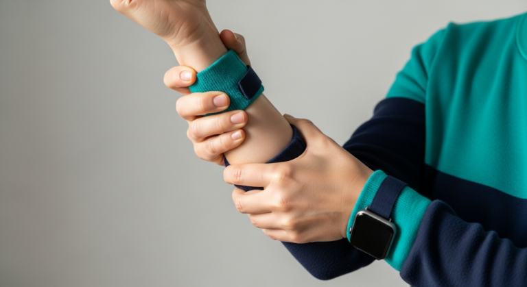
When Your Hand Doesn't Feel Like Your Own
It often starts subtly. A pins-and-needles sensation in the thumb that wakes you at 3 a.m. A dull ache along the wrist after a long day at the keyboard. A strange weakness when you try to open a jar or button a shirt.
For many people, wrist pain and hand numbness are dismissed as the inevitable cost of modern life — too much typing, too much scrolling, too much lifting a toddler. But when symptoms persist, escalate at night, or begin to interfere with the tasks that make up a normal day, the question quickly shifts from "Is this normal?" to "What is actually happening inside my wrist?"
The wrist and hand are astonishingly complex. Within a few centimetres sit nine flexor tendons, the median and ulnar nerves, a dense network of small joints, ligaments, and synovial spaces — any of which can become inflamed, compressed, or damaged. That anatomical density is why symptoms can feel confusingly similar even when the underlying causes are very different.
Carpal tunnel syndrome (CTS) is the most well-known culprit, and rightly so — it is the most common peripheral nerve entrapment worldwide. But it is far from the only explanation. A ganglion cyst, de Quervain's tenosynovitis, tendon inflammation, or early inflammatory arthritis can all masquerade as one another, particularly in the early stages when swelling is minimal and imaging is the clearest way to distinguish them.
This article is designed for the awareness stage of your journey: you are noticing symptoms, you are researching, and you want a clear, expert-led explanation of what may be happening — and when you need to act quickly.
---
⚠ Red Flags: When Wrist or Hand Symptoms Need Urgent Care
Most wrist pain is not an emergency. However, certain features warrant same-day assessment at A&E or an urgent GP appointment rather than a planned outpatient scan. Please seek immediate medical attention if you experience any of the following:
🚨 Go to A&E Now
- Sudden loss of hand or finger function following trauma — a fall onto an outstretched hand can cause a scaphoid fracture that may be invisible on initial X-ray.
- A visibly deformed wrist after injury, or bone protruding through the skin.
- Rapidly spreading redness, heat, or swelling, especially with fever — this may indicate septic arthritis or a deep hand-space infection, both of which are surgical emergencies.
- A red streak tracking up the forearm from a wound or puncture (ascending cellulitis or lymphangitis).
- Sudden, severe, unrelenting pain with a pale, cold, or blue-tinged hand — possible acute arterial occlusion or compartment syndrome.
- Complete numbness of the hand with inability to move the fingers, particularly after a crush injury or tight cast.
⚠️ Seek Urgent (Same-Week) Review
- Sudden inability to extend the thumb after wrist trauma or prolonged wrist activity — this may indicate rupture of the extensor pollicis longus tendon.
- Progressive weakness or wasting of the muscles at the base of the thumb (the thenar eminence) — a sign of advanced median nerve compression that risks becoming irreversible.
- Persistent swelling of a single joint with no clear cause, especially with morning stiffness lasting more than an hour (possible inflammatory arthritis).
- Symptoms following a suspected DVT risk event (recent surgery, immobilisation, malignancy) — although DVT is rare in the upper limb, it does occur and requires prompt Doppler assessment.
If none of these apply, your symptoms are almost certainly best investigated in a calm, planned setting — and this is precisely where diagnostic ultrasound excels.
Understanding the Anatomy in Plain English
Before we can make sense of what might be causing your symptoms, it helps to picture what actually lives inside your wrist. You don't need a medical degree — just a working mental map.
Imagine your wrist as a narrow tunnel of bone and ligament, roughly the width of your thumb. The floor and walls of this tunnel are formed by the small carpal bones, arranged in a shallow arch. The roof is a tough, fibrous band called the transverse carpal ligament (or flexor retinaculum), stretched tightly across the top like the lid of a shoebox.
Through this confined space pass ten structures: nine flexor tendons — the cables that curl your fingers and thumb — and one nerve. That nerve is the median nerve, and it is the star of this story.
What the Median Nerve Does
The median nerve is essentially the electrical cable that supplies sensation to the thumb, index finger, middle finger, and the thumb-side half of the ring finger. Notice which finger is missing: the little finger, which is served by the ulnar nerve. This distinction matters enormously for diagnosis, because tingling confined to the little finger points away from carpal tunnel syndrome and toward a different problem entirely (often ulnar nerve compression at the elbow).
The median nerve also powers several small muscles at the base of the thumb — the thenar muscles — which allow you to bring your thumb across your palm to touch your little finger. This movement, called opposition, is uniquely human and essential for grip. When the nerve is compressed for too long, these muscles waste away, and the fleshy pad at the base of the thumb visibly flattens.
Why the Tunnel Matters
Because the carpal tunnel is a closed, non-expandable space, anything that increases pressure inside it — swollen tendon sheaths, fluid retention, a ganglion cyst, thickened synovium from arthritis, or even pregnancy-related tissue changes — squeezes the median nerve first. The nerve is the softest, most vulnerable structure in the tunnel, and it responds to sustained pressure by first becoming irritable (tingling, burning) and eventually becoming damaged (numbness, weakness, muscle wasting).
Outside the tunnel, the wrist contains further structures that commonly cause pain:
- The first dorsal compartment on the thumb-side of the wrist, home to the tendons implicated in de Quervain's tenosynovitis.
- Small joint capsules and ligaments that can leak synovial fluid to form a ganglion cyst — the most common soft-tissue lump in the wrist.
- The scapholunate ligament, injury to which can cause deep, persistent wrist pain after a fall.
Understanding this layout is the key to understanding why one person's "wrist pain" is not the same as another's — and why imaging that can actually see these structures is so valuable.
---
Carpal Tunnel Syndrome: The Classic Picture
Carpal tunnel syndrome is the most common peripheral nerve entrapment condition in the world, affecting an estimated 3–5% of the general population and considerably more among people in repetitive-hand occupations, pregnant women, and those with diabetes, hypothyroidism, or rheumatoid arthritis (Padua et al., 2016).
Yet despite how common it is, CTS is frequently misidentified — either self-diagnosed when the true cause is something else, or dismissed as "just a bit of tingling" until nerve damage becomes advanced. The clinical picture is usually recognisable once you know what to look for.
The Hallmark Symptoms
Classical CTS follows a fairly predictable pattern:
- Nocturnal tingling and numbness in the thumb, index, middle, and radial half of the ring finger — the median nerve distribution. Patients typically describe waking in the early hours, shaking the hand vigorously to "get the feeling back." This shaking manoeuvre is so characteristic it has its own name: the flick sign.
- Symptoms triggered by sustained postures — holding a phone, driving, reading a book, or gripping a steering wheel. Anything that keeps the wrist flexed or extended for long periods raises pressure inside the tunnel.
- A burning or electric quality to the discomfort, distinct from the dull ache of tendon or joint pain.
- Clumsiness and dropping objects — as the nerve becomes more compromised, fine motor control and grip strength suffer. Patients often notice they can no longer feel the difference between a coin and a button in a pocket.
- Thenar muscle wasting in advanced cases — a visible hollowing at the base of the thumb, signalling that treatment should not be delayed.
Why Symptoms Are Worst at Night
Two mechanisms conspire against you when you sleep. First, most people flex or extend their wrists unconsciously while dreaming, dramatically raising pressure inside the tunnel. Second, fluid redistributes across the body when lying flat, subtly increasing tissue volume in the extremities. Together, these produce the classic 3 a.m. wake-up call that sends so many patients searching for answers.
The Role of Ultrasound in Diagnosis
Historically, CTS was diagnosed clinically and confirmed with nerve conduction studies (NCS) — a test involving small electrical shocks to measure how quickly signals travel along the median nerve. NCS remains valuable, but it has meaningful limitations: it can be uncomfortable, is not always available promptly, and — crucially — cannot show why the nerve is compressed.
High-resolution ultrasound has transformed the diagnostic pathway. By measuring the cross-sectional area (CSA) of the median nerve at the wrist, sonographers can quantify swelling of the nerve just before it enters the tunnel. A CSA above approximately 10 mm² is widely accepted as supportive of CTS, with higher values correlating with greater severity (Fowler et al., 2011; Cartwright et al., 2012).
More importantly, ultrasound answers a question NCS cannot: is there something structural inside the tunnel causing the compression? A ganglion cyst pressing on the nerve, an anomalous muscle belly, tenosynovitis of the flexor tendons, or a rare space-occupying lesion will all be visible in real time. This changes management — a nerve compressed by a cyst has a different treatment pathway than one compressed by generalised tendon swelling.
Ultrasound is also dynamic: we can watch the nerve as you move your fingers, assessing whether it glides normally or tethers abnormally against surrounding tissue. This is invaluable information that no static test can provide.
Beyond Carpal Tunnel: What Else Could It Be?
Carpal tunnel syndrome may be the most familiar diagnosis, but a significant proportion of patients who arrive convinced they have CTS turn out to have something else entirely — or CTS coexisting with a second condition. Recognising the alternatives is essential, because the treatment pathways diverge sharply.
Ganglion Cysts
A ganglion cyst is a fluid-filled swelling that arises from a joint capsule or tendon sheath, most commonly on the dorsal (back) surface of the wrist, though volar (palm-side) ganglia are also frequent and more likely to cause nerve symptoms.
The typical presentation is a firm, rounded lump that fluctuates in size — sometimes disappearing entirely before returning weeks later. Ganglia are filled with a viscous, jelly-like fluid derived from synovial tissue, and although they are entirely benign, they can cause discomfort by pressing on adjacent nerves or restricting joint movement.
The concern with volar ganglia is their proximity to the median nerve and radial artery. A small, deep ganglion sitting within or near the carpal tunnel can produce CTS-like symptoms without any visible lump at all. Ultrasound is exquisitely sensitive to these lesions, distinguishing a simple cyst from a solid mass in seconds and confirming its relationship to surrounding nerves and vessels — information that directly guides whether aspiration, surgical excision, or simple observation is appropriate.
De Quervain's Tenosynovitis
Named after the Swiss surgeon Fritz de Quervain, this condition involves inflammation of the tendon sheath containing the abductor pollicis longus and extensor pollicis brevis tendons — the two structures that run along the thumb-side of the wrist and control thumb movement.
The classic patient is a new parent (particularly a mother in the postpartum period), a keen gardener, or someone who has recently taken up a repetitive thumb-heavy activity such as texting, gaming, or racquet sports. The pain is sharply localised to the radial (thumb) side of the wrist, worsened by gripping, lifting, or wringing motions, and reliably reproduced by the Finkelstein test — tucking the thumb into the palm and gently ulnar-deviating the wrist.
Ultrasound reveals thickening of the tendon sheath, fluid within it, and hypervascularity on Doppler imaging — findings that not only confirm the diagnosis but also allow precise, image-guided corticosteroid injection into the sheath when conservative measures fail.
Flexor Tenosynovitis and Trigger Finger
Inflammation of the flexor tendon sheaths in the palm can cause stiffness, catching, or locking of a finger — the phenomenon known as trigger finger (stenosing tenosynovitis). Patients often feel a tender nodule in the palm and describe the finger "sticking" in flexion before releasing with a painful snap. Ultrasound clearly demonstrates the thickened A1 pulley and the associated tendon nodule, and can guide targeted injection therapy.
Early Inflammatory Arthritis
Perhaps the most important condition not to miss is early rheumatoid arthritis or another inflammatory arthropathy. Wrist involvement is common and often precedes changes visible on X-ray by months or even years.
Ultrasound can detect synovitis (inflamed joint lining), tenosynovitis, and early bone erosions long before radiographs turn positive, allowing rheumatologists to initiate disease-modifying treatment during the crucial "window of opportunity" that dramatically improves long-term outcomes (Wakefield et al., 2004).
Scapholunate Ligament Injury and Occult Fracture
Deep, persistent wrist pain following a fall — even a seemingly minor one — should always raise the possibility of a scapholunate ligament tear or an occult scaphoid fracture. These injuries are notoriously easy to miss on standard X-rays and can lead to progressive wrist arthritis if untreated. Ultrasound and, where necessary, MRI provide much more sensitive assessment.
---
From Our Practice
[SUGGESTED — verify with clinician]
At IUS London, one of the most consistent patterns we observe in our MSK ultrasound clinic is the patient who arrives with a firm self-diagnosis of carpal tunnel syndrome — often after weeks of online research and a GP consultation — only for imaging to reveal a rather different story.
A recurring scenario involves patients in their 30s to 50s, frequently working in desk-based or manual roles, who describe classic-sounding tingling in the hand alongside a dull ache on the thumb side of the wrist. They anticipate a straightforward measurement of the median nerve and confirmation of CTS. In a meaningful minority of these cases, we find two coexisting problems: a mildly enlarged median nerve (CSA around 10–12 mm²) and clear-cut de Quervain's tenosynovitis of the first dorsal compartment. The thumb-side pain, which the patient had attributed to "the carpal tunnel spreading," is in fact a distinct tendon problem requiring different management.
We also see a smaller but clinically important cohort — often postpartum women or patients with new-onset symptoms — in whom the CTS-like tingling turns out to be driven by a small volar ganglion cyst compressing the median nerve. This cyst is often too small to be palpable but is clearly visible on ultrasound, immediately changing the treatment plan from conservative management to targeted aspiration.
The operational lesson from our clinic is straightforward: wrist symptoms rarely have a single cause, and a scan that examines the whole wrist — not just the median nerve — consistently uncovers findings that reshape the treatment plan.
---
Comparing Imaging Modalities for Wrist Symptoms
Choosing the right investigation depends on what you and your clinician are trying to answer. Each modality has genuine strengths and honest limitations.
| Feature | **High-Resolution Ultrasound** | **MRI** | **X-ray** | **Nerve Conduction Studies (NCS)** |
|---|---|---|---|---|
| **Best for** | Median nerve, tendons, ganglia, dynamic assessment, synovitis | Ligaments, cartilage, deep bone marrow, occult fractures | Bony alignment, established arthritis, fractures | Quantifying nerve conduction velocity |
| **Median nerve CSA measurement** | ✅ Excellent (real-time, quantitative) | ⚠️ Possible but less practical | ❌ Not visible | ❌ Assesses function, not anatomy |
| **Detects ganglion cysts** | ✅ Excellent | ✅ Excellent | ❌ Rarely visible | ❌ No |
| **De Quervain's / tenosynovitis** | ✅ Excellent with Doppler | ✅ Good | ❌ No | ❌ No |
| **Early synovitis / erosions** | ✅ Very good | ✅ Excellent (gold standard) | ⚠️ Only in later disease | ❌ No |
| **Dynamic (movement) assessment** | ✅ Unique strength | ❌ Static only | ❌ Static only | ⚠️ Partial |
| **Guides injection therapy** | ✅ Real-time guidance | ❌ No | ❌ No | ❌ No |
| **Patient comfort** | ✅ No enclosure, no contrast | ⚠️ Enclosed, longer scan | ✅ Quick | ⚠️ Mild electrical discomfort |
| **Radiation** | ✅ None | ✅ None | ⚠️ Low dose | ✅ None |
| **Typical wait time (private)** | Days | 1–2 weeks | Days | Weeks |
| **Cost profile** | £ | £££ | £ | ££ |
For the majority of patients presenting with wrist pain or hand numbness at the awareness stage of their journey, ultrasound is the ideal first-line investigation: it directly visualises the structures most likely to be responsible, quantifies median nerve involvement, identifies coexisting pathology, and can transition seamlessly into image-guided treatment if appropriate. MRI remains invaluable for deeper ligamentous or cartilaginous questions, and X-ray retains its role after significant trauma or in established arthritis.
What to Expect at Your Ultrasound Appointment
Many patients tell us that the uncertainty surrounding a scan is more stressful than the scan itself. In reality, a wrist and hand ultrasound at IUS London is one of the most comfortable and least invasive investigations in medicine.
Before the Scan
You will be greeted by a consultant sonographer or musculoskeletal radiologist who will take a focused clinical history — when the symptoms began, what makes them better or worse, whether they wake you at night, and what specific movements or tasks trigger them. This conversation genuinely shapes the scan: a patient describing thumb-side pain will receive a different scanning protocol than one describing purely nocturnal tingling.
You will be asked to remove any watches, bracelets, or rings from the affected side. There is no need to change into a gown — a rolled-up sleeve is usually sufficient.
During the Scan
You will sit comfortably opposite the sonographer, with your forearm resting palm-up on a cushioned pad. A small amount of warmed water-based gel is applied to the skin, and a handheld probe is moved gently across the wrist and hand.
The scan is entirely painless. You may feel light pressure as the sonographer angles the probe to obtain optimal images, but nothing sharp or uncomfortable. Expect the following elements:
- Static imaging of the median nerve, with measurement of its cross-sectional area at the level of the pisiform bone.
- Dynamic imaging, where you will be asked to flex and extend the fingers so the sonographer can observe tendon glide and nerve mobility.
- Doppler assessment, which uses colour overlay to highlight inflammation or hypervascularity within tendon sheaths or joint linings.
- Comparative scanning of the opposite (asymptomatic) wrist, which is often invaluable for context.
The examination typically takes 20 to 30 minutes.
After the Scan
You will receive a verbal explanation of the findings immediately, often with the images visible on screen so you can see exactly what has been identified. A detailed written report follows, which can be shared with your GP, physiotherapist, or hand surgeon. If image-guided injection is appropriate and you wish to proceed, it can frequently be arranged at a subsequent visit.
---
Practical Steps Before Your Appointment
A little preparation makes a meaningful difference to the quality of your consultation and the specificity of your report.
Track Your Symptoms
For the week before your scan, keep a brief note of:
- When symptoms occur — night, morning, during specific tasks.
- Which fingers are involved — this is the single most useful clue for nerve-related diagnoses.
- What relieves them — shaking the hand, changing position, resting.
- Any functional impact — dropping objects, difficulty with buttons or keys, weakened grip.
Bring Relevant Documents
If you have had previous imaging, nerve conduction studies, blood tests, or a referral letter, bring copies (digital or paper). Even old reports can help contextualise new findings.
Avoid Splint Removal Just Before the Scan
If you routinely wear a wrist splint, wear it as normal on the day. There is no need to leave it off — we simply remove it during the examination.
Skip Hand Cream on the Day
Moisturisers and oils can interfere with ultrasound gel adherence. A clean, dry forearm and hand are ideal.
Prepare Your Questions
Awareness-stage patients often want to leave the appointment with clarity on three things: what is causing my symptoms, how serious is it, and what should I do next? Writing your questions down in advance ensures nothing is missed in the moment.
---
When to Book an Ultrasound
Not every twinge warrants imaging. However, the following situations are strong indications that a private MSK ultrasound will meaningfully shorten your path to answers:
- Nocturnal tingling in the thumb, index, middle, or ring finger persisting for more than two to four weeks.
- A visible or palpable lump on the wrist, whether painful or painless.
- Localised thumb-side wrist pain worsened by gripping or lifting, particularly in new parents or after a change in activity.
- Persistent wrist pain following a fall, even if an X-ray was reported as normal.
- Symptoms that have not resolved after a course of splinting, activity modification, or physiotherapy.
- Early signs of muscle wasting at the base of the thumb — a signal that intervention should not be delayed.
- Suspected inflammatory arthritis, particularly with morning stiffness or symmetrical joint symptoms.
- A need for image-guided injection rather than a "blind" procedure.
Booking privately means you can typically be scanned within a few working days, receive same-visit results, and move directly into a treatment conversation rather than waiting weeks for sequential appointments through separate services.
---
Take the Next Step Toward Clarity
Wrist pain and hand numbness are among the most disruptive symptoms a person can experience — not because they are dangerous in most cases, but because the hand is so central to how we work, care for our families, and express ourselves. Living with uncertainty about what is happening inside your wrist takes a genuine toll.
A focused, consultant-led musculoskeletal ultrasound scan gives you something that no amount of online research can: a direct look at the tissues causing your symptoms, and a clear, personalised explanation of what to do about them.
At IUS London, our MSK ultrasound service is designed around this principle — same-day reporting, expert scanning by consultants who scan wrists every week, and a seamless pathway into treatment when it is needed.
👉 [Book a Private MSK Ultrasound Scan at IUS London](/services/msk-ultrasound-scan/)
If you are unsure whether ultrasound is the right investigation for your specific symptoms, our clinical team is happy to advise before you book. Clarity is the first step toward relief — and it is closer than you think.
---
References
1. Padua, L., Coraci, D., Erra, C., Pazzaglia, C., Paolasso, I., Loreti, C., Caliandro, P., & Hobson-Webb, L. D. (2016). Carpal tunnel syndrome: clinical features, diagnosis, and management. The Lancet Neurology, 15(12), 1273–1284. https://doi.org/10.1016/S1474-4422(16)30231-9
2. Fowler, J. R., Gaughan, J. P., & Ilyas, A. M. (2011). The sensitivity and specificity of ultrasound for the diagnosis of carpal tunnel syndrome: a meta-analysis. Clinical Orthopaedics and Related Research, 469(4), 1089–1094. https://doi.org/10.1007/s11999-010-1637-5
3. Cartwright, M. S., Hobson-Webb, L. D., Boon, A. J., Alter, K. E., Hunt, C. H., Flores, V. H., Werner, R. A., Shook, S. J., Thomas, T. D., Primack, S. J., & Walker, F. O. (2012). Evidence-based guideline: neuromuscular ultrasound for the diagnosis of carpal tunnel syndrome. Muscle & Nerve, 46(2), 287–293. https://doi.org/10.1002/mus.23389
4. Wakefield, R. J., Green, M. J., Marzo-Ortega, H., Conaghan, P. G., Gibbon, W. W., McGonagle, D., Proudman, S., & Emery, P. (2004). Should oligoarthritis be reclassified? Ultrasound reveals a high prevalence of subclinical disease. Annals of the Rheumatic Diseases, 63(4), 382–385. https://doi.org/10.1136/ard.2003.007062
5. McDonagh, C., Alexander, M., & Kane, D. (2015). The role of ultrasound in the diagnosis and management of carpal tunnel syndrome: a new paradigm. Rheumatology, 54(1), 9–19. https://doi.org/10.1093/rheumatology/keu275
Book Your Private MSK Ultrasound Scan in London
Same-day appointments available in Kensington. No GP referral required. HCPC-registered consultant sonographers.