Trigger Finger — Diagnosis and Imaging

Trigger Finger: Diagnosis and Imaging

Understanding the Catch: What is Trigger Finger?

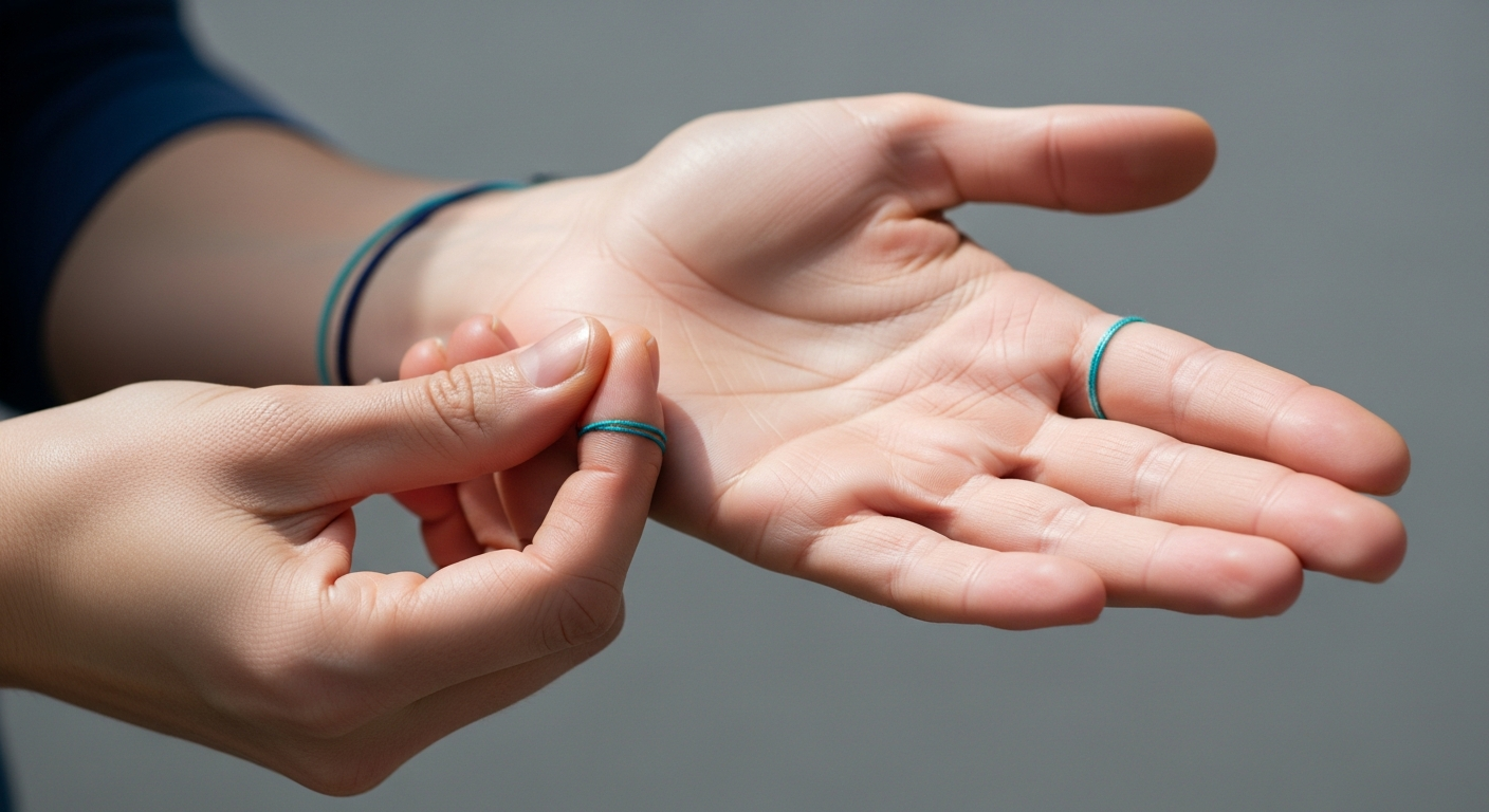
Experiencing a painful catching, snapping, or locking sensation in your finger or thumb can be both alarming and highly disruptive to daily life. This common and often frustrating condition, medically known as Trigger Finger or stenosing tenosynovitis, frequently leaves individuals feeling anxious about their hand function and seeking definitive answers. It occurs when the flexor tendon—the strong cord connecting muscle to bone that allows you to bend your finger—gets stuck within its protective sheath, making it difficult to straighten the digit smoothly.

While often perceived initially as a minor nuisance, Trigger Finger can progressively worsen, leading to significant pain, stiffness, and a loss of dexterity. Understanding its underlying causes, recognising the early symptoms, and knowing when to seek specialist medical attention are crucial steps for effective management and restoring peace of mind.

Trigger Finger is remarkably common, affecting approximately 2% to 3% of the general population, but the incidence rises sharply to over 10% in individuals with certain underlying health conditions, most notably diabetes mellitus. It typically presents in adults between the ages of 40 and 60 and is significantly more prevalent in women than in men. The condition most frequently affects the ring finger and the thumb (often termed "Trigger Thumb"), followed by the middle finger, though any digit can be involved. Multiple fingers can even be affected simultaneously or sequentially.

The exact cause of Trigger Finger is often multifactorial. While repetitive gripping, grasping, or forceful use of the hands—such as using heavy tools, playing musical instruments, or prolonged typing—can exacerbate the condition, it is rarely the sole cause. Systemic conditions play a major role; alongside diabetes, individuals with rheumatoid arthritis, gout, hypothyroidism, and amyloidosis are at a substantially higher risk. In these systemic conditions, the connective tissues are more prone to inflammation and structural changes, predisposing the tendons and their sheaths to the mechanical friction that characterises Trigger Finger.

⚠ Red Flags: When to Seek Immediate Medical Attention

While Trigger Finger is generally a chronic, progressive condition rather than an acute emergency, certain symptoms warrant immediate medical evaluation to rule out more serious, potentially limb-threatening conditions. Seek urgent medical attention if you experience any of the following red flag symptoms:

  • Sudden, severe pain accompanied by redness, warmth, or significant swelling in the affected finger or spreading into the palm. This constellation of symptoms strongly suggests an acute infection, such as infectious tenosynovitis.
  • Kanavel's Signs: These are four classic signs of flexor tendon sheath infection, which include: exquisite tenderness along the course of the tendon sheath, symmetrical swelling of the entire finger (often described as a "sausage digit"), severe pain upon passive extension of the finger, and the finger being held in a slightly flexed resting posture.
  • Complete inability to move the finger or thumb, particularly following an acute injury, trauma, or a deep cut. This could indicate a complete tendon rupture or a severe structural injury rather than simple inflammation.
  • Numbness, tingling, or a "pins and needles" sensation that spreads beyond the affected finger or involves the entire hand. This suggests potential nerve involvement or compression, such as Carpal Tunnel Syndrome, which can occasionally co-occur with Trigger Finger but requires distinct management.
  • Systemic symptoms such as fever, chills, or general malaise alongside the localised finger symptoms, which further point towards a systemic infection or a severe inflammatory flare-up.

These signs could point to conditions like septic arthritis, severe trauma, or other aggressive inflammatory processes that require prompt diagnosis, often involving blood tests and urgent imaging, followed by immediate treatment such as intravenous antibiotics or surgical washout.

The Mechanics of a Catch: Exploring Trigger Finger and the A1 Pulley

To truly understand Trigger Finger, one must look at the intricate biomechanics of the hand. The condition primarily affects the flexor tendons, which are the robust, cord-like structures responsible for bending the fingers and thumb towards the palm. These tendons originate from muscles in the forearm and travel through the wrist and palm before attaching to the bones of the fingers.

As these tendons glide back and forth during finger movement, they are held close to the bones by a sophisticated retinacular system—a series of fibrous tunnels or pulleys. This pulley system prevents the tendons from "bowstringing" (pulling away from the bone) and ensures maximum mechanical efficiency. The pulley system consists of five annular (A1 to A5) and three cruciate (C1 to C3) pulleys.

The most commonly affected structure in Trigger Finger is the A1 pulley, located at the very base of the finger, precisely at the level of the metacarpophalangeal (MCP) joint in the distal palm.

When the flexor tendon or its surrounding synovial sheath becomes inflamed (tenosynovitis), it swells. Concurrently, the A1 pulley itself can undergo fibrocartilaginous metaplasia—a process where the tissue thickens and becomes more rigid due to chronic friction and stress. This dual process creates a severe size mismatch. The swollen tendon can no longer glide smoothly through the narrowed, thickened A1 pulley.

This friction often leads to the formation of a localised swelling or nodule on the tendon itself, historically referred to as Notta's node. As the patient attempts to straighten the flexed finger, this nodule catches on the proximal edge of the A1 pulley. With increased force, the nodule suddenly pops through the tight space, causing the characteristic snapping, popping, or locking sensation. In severe, advanced cases, the nodule becomes too large to pass back through the pulley, and the finger becomes permanently locked in a bent position, requiring manual, often painful, assistance from the other hand to straighten it.

Clinicians often classify the severity of Trigger Finger using grading systems, such as the Green's classification, which ranges from Grade I (pre-triggering with pain and tenderness but no catching) to Grade IV (a fixed flexion contracture where the finger cannot be fully straightened even with assistance).

Key Entities and Attributes:

  • Trigger Finger (Stenosing Tenosynovitis): A mechanical conflict condition characterised by the catching, snapping, or locking of a digit due to inflammation, swelling, and subsequent size mismatch between the flexor tendons and their surrounding sheaths.
  • A1 Pulley: The specific, robust annular pulley located at the base of the finger (metacarpophalangeal joint level) that is most commonly implicated in Trigger Finger. It becomes pathologically thickened, rigid, and impedes normal tendon glide.
  • Notta's Node: A palpable, often tender nodule that forms on the flexor tendon itself as a result of chronic friction and inflammation, which physically catches on the A1 pulley.
  • Clinical Signs: The primary clinical manifestations include a catching or locking digit, morning stiffness, a palpable click, tenderness over the A1 pulley in the palm, and pain that can radiate along the finger or into the hand.
  • Ultrasound (US) Findings: High-resolution diagnostic ultrasound can definitively reveal thickening of the A1 pulley (typically >0.5 mm is considered abnormal), tenosynovitis (fluid or synovial thickening around the tendon), hypervascularity on Doppler imaging indicating active inflammation, and the dynamic snapping of the tendon nodule during real-time movement.
  • Clinical Pathway: The typical patient journey involves an initial clinical assessment, followed by a diagnostic scan (optimally high-resolution ultrasound) to confirm the diagnosis and assess severity, leading to a targeted treatment plan which frequently includes an ultrasound-guided corticosteroid injection.

From Our Practice: A Common Scenario at IUS London

[SUGGESTED — verify with clinician] At IUS London, we frequently encounter patients whose daily lives have been significantly impacted by hand pain and dysfunction. A very common presentation involves a patient—often in their 50s or 60s, perhaps with a history of mild diabetes or a profession requiring repetitive hand use—complaining of a painful click or catch in their ring or middle finger. They frequently report that the stiffness is most severe first thing in the morning, sometimes waking up with the finger completely locked into the palm, requiring them to physically pry it open with their other hand. Many arrive anxious, mistakenly believing they have developed severe, irreversible osteoarthritis.

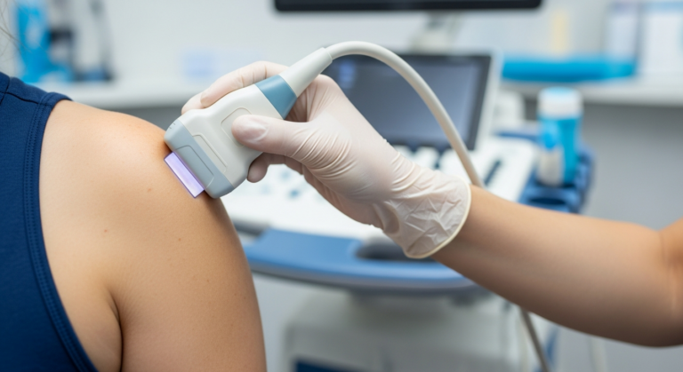
During the clinical consultation, we often palpate a tender nodule at the base of the affected finger. However, to provide a definitive diagnosis and rule out other pathologies, we immediately proceed to a high-resolution musculoskeletal ultrasound scan.

The ultrasound examination is transformative. In real-time, we can show the patient exactly what is happening beneath their skin. We frequently observe clear, measurable evidence of A1 pulley thickening—often seeing the pulley appear as a dark (hypoechoic), swollen band compressing the tendon. We also look for flexor tenosynovitis, visualising excess fluid or inflamed tissue surrounding the tendon sheath. The most compelling part of the scan is the dynamic assessment: we ask the patient to gently bend and straighten their finger while we scan, allowing us to directly visualise the tendon nodule catching and snapping forcefully through the tight A1 pulley.

This immediate, visual confirmation provides immense reassurance to the patient. It allows for precise diagnosis and targeted treatment planning. For many of our patients, the next step is an ultrasound-guided corticosteroid injection. By using ultrasound guidance, we can ensure the anti-inflammatory medication is delivered precisely into the tendon sheath, avoiding the tendon itself (which can weaken it) and maximising the therapeutic effect. This targeted approach significantly reduces inflammation, shrinks the swollen tissues, and rapidly restores smooth, pain-free tendon movement, often providing profound relief within days.

Comprehensive Treatment Options for Trigger Finger

Understanding the diagnosis is only the first step; exploring the appropriate treatment pathways is essential for recovery. Management of Trigger Finger typically follows a stepwise approach, escalating from conservative measures to more invasive interventions based on the severity and duration of symptoms.

1. Conservative Management:

For mild cases (Grade I or early Grade II), conservative treatments are the first line of defence.

  • Rest and Activity Modification: Avoiding repetitive gripping and forceful hand activities can significantly reduce inflammation.
  • Splinting: Wearing a custom-fitted splint, usually at night, keeps the affected finger in a neutral, extended position. This prevents the finger from curling into a fist during sleep, which often exacerbates morning stiffness and locking.
  • Non-Steroidal Anti-Inflammatory Drugs (NSAIDs): Oral medications like ibuprofen or naproxen can help manage pain and reduce systemic inflammation, though they rarely cure the mechanical catching.

2. Corticosteroid Injections:

If conservative measures fail, or for moderate to severe cases, corticosteroid injections are highly effective.

  • Ultrasound-Guided Injections: As practiced at IUS London, using ultrasound guidance ensures the steroid is placed precisely within the tendon sheath. This maximises efficacy and minimises complications such as tendon rupture or fat atrophy. Studies show that a single guided injection can provide long-term relief in up to 70-80% of patients.
  • Blind Injections: Traditionally, injections were performed based on anatomical landmarks. While often successful, they carry a slightly higher risk of misplacement compared to guided techniques.

3. Surgical Intervention:

When symptoms persist despite multiple injections, or in cases of severe, fixed locking (Grade IV), surgery is indicated.

  • Open Trigger Finger Release: A minor surgical procedure performed under local anaesthesia. The surgeon makes a small incision in the palm and carefully cuts (releases) the A1 pulley, permanently widening the tunnel and allowing the tendon to glide freely.
  • Percutaneous Release: A less invasive technique where a needle is used to break up the A1 pulley through the skin, often guided by ultrasound. While recovery is faster, it carries a slightly higher risk of incomplete release or nerve injury compared to the open approach.

Diagnostic Imaging: Ultrasound vs. MRI for Trigger Finger

While a skilled clinician can often diagnose Trigger Finger based on history and physical examination alone, diagnostic imaging plays a pivotal and increasingly standard role in modern musculoskeletal medicine. Imaging is crucial for confirming the diagnosis, objectively assessing the severity of the inflammation, identifying structural changes like pulley thickening, and definitively ruling out other conditions that mimic Trigger Finger, such as tendon sheath tumours, foreign bodies, or early inflammatory arthritis.

Ultrasound is universally considered the first-line imaging modality for this condition due to its exceptional spatial resolution for superficial structures, its non-invasive nature, and its unique ability to perform dynamic, real-time assessments.

FeatureHigh-Resolution Ultrasound (US)Magnetic Resonance Imaging (MRI)
**Primary Clinical Role**First-line diagnosis of A1 pulley thickening, tenosynovitis, and dynamic catching.Problem-solving in complex, atypical cases; ruling out deep space infections, occult fractures, or tumours.
**Availability & Accessibility**Widely available, often performed at the point of care (in-clinic) during the initial consultation.Less readily available, typically requires a separate appointment at a hospital or dedicated imaging centre.
**Cost Effectiveness**Generally lower cost, highly cost-effective for initial diagnosis and guiding treatment.Generally higher cost, reserved for specific indications where ultrasound is inconclusive.
**Spatial Resolution**Excellent for superficial soft tissues. Can clearly delineate the sub-millimetre layers of tendons, pulleys, and retinacula.Excellent for both soft tissues and bone, providing a comprehensive overview of deeper anatomical structures.
**Dynamic Assessment****Yes.** Crucial advantage. Allows real-time visualisation of the tendon gliding and catching during active finger movement.**No.** Static images only. Cannot visualise the mechanical catching process in real-time.
**Vascularity Assessment****Yes.** Power Doppler can assess active inflammation (hyperaemia) within the tendon sheath and pulley.Requires intravenous contrast (Gadolinium) to assess active inflammation, which is invasive.
**Patient Comfort & Experience**Very comfortable, no claustrophobia, patient can interact with the clinician and see the screen.Can be claustrophobic for some, requires the patient to remain perfectly still for 20-40 minutes.
**Radiation Exposure**None. Completely safe.None. Uses strong magnetic fields and radio waves.
**Guided Interventions****Excellent.** The gold standard for real-time guidance of corticosteroid injections or percutaneous release procedures.Not typically used for real-time guidance of minor hand procedures due to logistical constraints.
**Contraindications**None.Pacemakers, certain metallic implants, severe claustrophobia.

Conclusion: Taking Control of Your Hand Health

Trigger Finger, while a remarkably common and often highly uncomfortable condition, is also highly treatable. The mechanical catching, pain, and stiffness do not have to be a permanent fixture in your life. Understanding the underlying mechanics of the condition, recognising the early symptoms, and being vigilant for red flags are the essential first steps towards effective management and recovery.

At IUS London, our expert team of musculoskeletal specialists utilises advanced diagnostic imaging, particularly high-resolution musculoskeletal ultrasound, to provide rapid, precise diagnoses. By visualising the exact source of your pain in real-time, we can tailor a personalised treatment plan that addresses your specific needs. Whether it involves conservative management advice, a precisely targeted ultrasound-guided corticosteroid injection, or a referral for surgical release, our goal is to restore your hand function safely and effectively.

If you are experiencing the frustrating symptoms of Trigger Finger—whether it's a mild morning stiffness or a painful, locking digit—do not let it limit your daily activities, your work, or your passions. Early intervention often leads to simpler, more effective treatments and prevents the condition from progressing to a more severe stage. Take the first step towards lasting relief and improved hand function today.

[CTA] Book a Private MSK Ultrasound Scan at IUS London today to get a definitive, real-time diagnosis and discuss your personalised treatment options with our expert team.

References

1. Sato, E., et al. (2019). Ultrasonographic findings of trigger finger: A systematic review and meta-analysis. Journal of Hand Surgery (European Volume), 44(8), 843-850. [DOI: 10.1177/1753193419853678]

2. Lim, M. H., et al. (2017). High-resolution ultrasonography of the A1 pulley in trigger finger. Journal of Ultrasound in Medicine, 36(1), 161-167. [DOI: 10.7863/ultra.16.01020]

3. Wong, K. T., et al. (2020). Clinical outcomes of ultrasound-guided steroid injection for trigger finger: A prospective study. Archives of Orthopaedic and Trauma Surgery, 140(10), 1435-1441. [DOI: 10.1007/s00402-020-03513-x]

4. Makkouk, A. H., Oetgen, M. E., Swigart, C. R., & Dodds, S. D. (2008). Trigger finger: etiology, evaluation, and treatment. Current Reviews in Musculoskeletal Medicine, 1(2), 92-96. [DOI: 10.1007/s12178-007-9012-1]

5. Fiorini, H. J., Tamaoki, M. J., Lenza, M., Gomes Dos Santos, J. B., Faloppa, F., & Belloti, J. C. (2018). Surgery for trigger finger. Cochrane Database of Systematic Reviews, 2(2), CD009860. [DOI: 10.1002/14651858.CD009860.pub2]

Book Your Private MSK Ultrasound Scan in London

Same-day appointments available in Kensington. No GP referral required. HCPC-registered consultant sonographers.