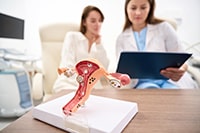
Why Am I Not Getting Pregnant? Understanding Fertility and When to Scan

Why Am I Not Getting Pregnant?

Understanding Infertility: A Comprehensive Guide

Infertility can be a challenging and emotionally taxing journey for many individuals and couples. Defined as the inability to conceive after a year of regular, unprotected intercourse (or six months for women over 35), it affects a significant portion of the population worldwide. This comprehensive guide aims to shed light on the various causes of infertility, the diagnostic process, and the available treatment options, providing a supportive resource for those navigating this complex landscape.

Common Causes of Infertility

Infertility is a complex condition with a multitude of potential causes, often involving a combination of factors from both partners. Understanding these underlying issues is the first step towards effective diagnosis and treatment. The primary categories of infertility causes include ovulatory disorders, tubal factors, male factor infertility, and unexplained infertility.

Anovulation: Disruptions in Ovulation

Anovulation, the absence of ovulation, is a common cause of female infertility, accounting for approximately 25% of all cases [1]. Ovulation is the process where a mature egg is released from the ovary, a crucial step for conception. When this process is irregular or absent, natural conception becomes difficult or impossible.

#### Polycystic Ovary Syndrome (PCOS)

Polycystic Ovary Syndrome (PCOS) is the most prevalent endocrine disorder among women of reproductive age, affecting approximately 5-10% of women globally, and is a leading cause of anovulation [5]. It is characterized by a constellation of symptoms including irregular or absent menstrual periods (oligomenorrhea or amenorrhea), elevated androgen levels (hyperandrogenism, leading to clinical signs like hirsutism, acne, and androgenic alopecia), and polycystic ovaries visible on ultrasound (presence of 12 or more follicles measuring 2-9 mm in diameter in at least one ovary, and/or increased ovarian volume >10 mL). The diagnostic criteria often follow the Rotterdam criteria, requiring at least two out of these three features after exclusion of other etiologies [6].

The pathophysiology of PCOS is complex and multifactorial, involving a intricate interplay of genetic predisposition, environmental factors, and endocrine dysregulation. A central feature is insulin resistance, which affects approximately 50-70% of women with PCOS, independent of obesity [7]. This resistance leads to compensatory hyperinsulinemia, which in turn stimulates ovarian androgen production and reduces hepatic synthesis of sex hormone-binding globulin (SHBG), further increasing free androgen levels. Elevated androgens disrupt the delicate hormonal balance required for normal follicular development. Specifically, they interfere with the maturation of ovarian follicles, leading to an accumulation of small, immature follicles that fail to ovulate, resulting in chronic anovulation. The excess androgens also contribute to the clinical manifestations such as hirsutism and acne. Furthermore, the altered gonadotropin secretion, with a relatively higher luteinizing hormone (LH) to follicle-stimulating hormone (FSH) ratio, is often observed in PCOS, which further exacerbates ovarian androgen production and impairs follicular growth.

#### Hypothalamic Amenorrhea

From an anatomical and physiological perspective, the hypothalamus, located at the base of the brain, acts as the central command center for reproductive function. It secretes GnRH in a pulsatile manner, which is critical for stimulating the anterior pituitary gland to release FSH and LH. FSH promotes the growth and maturation of ovarian follicles, while LH triggers ovulation. In hypothalamic amenorrhea, chronic energy deficit (due to low caloric intake and/or high energy expenditure) or psychological stress leads to a reduction in GnRH pulse frequency and amplitude. This diminished GnRH signaling results in decreased FSH and LH secretion, subsequently leading to inadequate follicular development and anovulation. The ovaries, lacking sufficient gonadotropic stimulation, become quiescent, leading to low estrogen levels and the absence of menstrual periods. This state is often reversible with lifestyle modifications, such as increasing caloric intake, reducing exercise intensity, and managing stress.

#### Premature Ovarian Insufficiency (POI)

Premature Ovarian Insufficiency (POI), sometimes referred to as premature ovarian failure, occurs when a woman's ovaries stop functioning normally before the age of 40. This results in irregular or absent periods and reduced fertility due to a depletion of ovarian follicles. POI can be caused by genetic factors, autoimmune diseases, chemotherapy, radiation therapy, or unknown reasons. Unlike menopause, POI can sometimes be intermittent, and spontaneous pregnancies can occur, though rarely.

Clinically, POI is diagnosed by the presence of amenorrhea for at least four months and elevated FSH levels (typically >25 mIU/mL) on two occasions more than four weeks apart, in women under 40 years of age [8]. The underlying pathology involves a significant reduction in the number of primordial follicles within the ovaries, or dysfunction of the remaining follicles. Genetic causes can include chromosomal abnormalities like Turner syndrome (45,X) or fragile X premutation. Autoimmune conditions, where the body's immune system mistakenly attacks ovarian tissue, are also a significant contributor. Environmental factors, such as exposure to toxins, and iatrogenic causes, like chemotherapy or radiation for cancer treatment, can also induce ovarian damage and follicle loss. The consequence of this follicular depletion or dysfunction is a failure of the ovaries to produce adequate estrogen and to release eggs regularly, leading to infertility. While spontaneous pregnancies are rare, the possibility underscores the difference between POI and true menopause, where ovarian function has completely ceased.

Tubal Factors: Blocked or Damaged Fallopian Tubes

Tubal factor infertility accounts for about 30% of female infertility cases and refers to conditions where the fallopian tubes are blocked or damaged, preventing the egg from reaching the uterus or sperm from reaching the egg [2]. The fallopian tubes are delicate structures, and their patency and function are crucial for natural conception.

#### Pelvic Inflammatory Disease (PID)

Pelvic Inflammatory Disease (PID) is a common and serious infection of the female reproductive organs, including the uterus, fallopian tubes, and ovaries. It is most frequently caused by sexually transmitted bacteria, particularly Chlamydia trachomatis and Neisseria gonorrhoeae, which ascend from the vagina and cervix into the upper reproductive tract [9]. The inflammatory response triggered by these infections can lead to significant and often irreversible damage to the delicate ciliated epithelium lining the fallopian tubes. This damage can result in hydrosalpinx (fluid accumulation within the tube), pyosalpinx (pus accumulation), or complete occlusion of the tubal lumen. The cilia, microscopic hair-like structures, are crucial for propelling the egg from the ovary towards the uterus. When these are damaged or destroyed, the egg's transport is severely compromised, increasing the risk of both infertility and ectopic pregnancy. Even subclinical or asymptomatic PID can cause significant tubal damage, highlighting the importance of early diagnosis and treatment of STIs.

#### Endometriosis

Endometriosis is a chronic, inflammatory condition characterized by the presence of endometrial-like tissue outside the uterus. This ectopic tissue, commonly found on the ovaries, fallopian tubes, peritoneum, and bowel, responds to hormonal fluctuations of the menstrual cycle, leading to cyclical bleeding, inflammation, and pain [10]. The presence of endometriosis can impair fertility through several mechanisms. Anatomically, the inflammatory process can lead to the formation of adhesions – fibrous bands of scar tissue that can distort the normal pelvic anatomy, tethering organs together and obstructing the fallopian tubes. This physical obstruction can prevent the egg from being captured by the fimbriae of the fallopian tube or from traveling down the tube to meet sperm. Physiologically, endometriosis creates a hostile peritoneal environment characterized by increased levels of inflammatory cytokines, prostaglandins, and macrophages. These factors can negatively impact oocyte quality, sperm function, fertilization, embryo development, and implantation, even in the absence of overt anatomical distortion [11]. Ovarian endometriomas (cysts on the ovaries) can also reduce ovarian reserve and interfere with normal follicular development.

#### Previous Abdominal Surgery

Any prior abdominal or pelvic surgery carries a risk of post-surgical adhesion formation, which can significantly impact fertility. Adhesions are bands of scar tissue that form between organs or between organs and the abdominal wall, often as a result of the body's healing response to surgical trauma. Common surgeries that can lead to fertility-impairing adhesions include appendectomy, C-sections, myomectomy (removal of uterine fibroids), and surgeries for ovarian cysts or endometriosis. These adhesions can physically distort the delicate anatomy of the fallopian tubes and ovaries, preventing the normal interaction between the egg and sperm. For instance, an adhesion might kink a fallopian tube, making it impassable, or encapsulate an ovary, preventing egg release. While modern surgical techniques aim to minimize adhesion formation, it remains a potential complication that can contribute to tubal factor infertility.

Male Factor Infertility

Male factor infertility contributes to approximately 30-50% of infertility cases, either as a sole cause or in combination with female factors [3]. It involves issues with sperm production, function, or delivery.

#### Sperm Production Disorders

Sperm production, or spermatogenesis, is a highly complex and tightly regulated process occurring within the seminiferous tubules of the testes. Disorders in this process can lead to a spectrum of issues, from azoospermia (complete absence of sperm in the ejaculate) to oligospermia (low sperm count) or asthenozoospermia (poor sperm motility). The underlying pathology can be broadly categorized into pre-testicular, testicular, and post-testicular causes.

  • Pre-testicular causes involve hormonal imbalances that affect testicular function. For example, hypogonadotropic hypogonadism results from insufficient production of GnRH, FSH, or LH by the hypothalamus or pituitary gland, leading to inadequate stimulation of the testes. Genetic conditions like Klinefelter syndrome (47,XXY karyotype) are characterized by testicular dysgenesis, leading to small, firm testes and impaired spermatogenesis. Other factors include systemic illnesses, certain medications, and lifestyle choices.
  • Testicular causes directly affect the testes' ability to produce sperm. Varicocele, an abnormal enlargement of veins within the scrotum, is a common correctable cause of male infertility, believed to impair spermatogenesis through increased scrotal temperature and oxidative stress [12]. Infections such as mumps orchitis (inflammation of the testes due to mumps virus) can cause irreversible damage to the seminiferous tubules. Cryptorchidism (undescended testes) and testicular trauma can also lead to impaired sperm production. Exposure to environmental toxins, radiation, or certain chemotherapy drugs can also directly damage germ cells.

#### Sperm Function Issues

Beyond mere numbers, the quality and functional capacity of sperm are paramount for successful fertilization. Issues with sperm morphology (abnormal shape), motility (progressive movement), or capacitation (the physiological changes sperm undergo in the female reproductive tract to become capable of fertilization) can significantly impair fertility, even if sperm count is normal. Teratozoospermia, characterized by a high percentage of abnormally shaped sperm, can hinder their ability to penetrate the egg's outer layers. Asthenozoospermia, poor sperm motility, means sperm may not be able to reach the egg in the fallopian tube. These functional deficits can stem from various pathologies:

  • Genetic defects: Subtle genetic abnormalities, not always detectable by standard karyotyping, can affect sperm development and function. For instance, microdeletions on the Y chromosome can lead to severe oligozoospermia or azoospermia.
  • Oxidative stress: An imbalance between the production of reactive oxygen species (ROS) and the body's antioxidant defenses can damage sperm DNA, membranes, and proteins, leading to impaired motility and function [13]. Sources of oxidative stress include infection, inflammation, varicocele, and lifestyle factors.
  • Immunological factors: In some men, the immune system mistakenly produces anti-sperm antibodies. These antibodies can bind to sperm, impairing their motility, ability to penetrate cervical mucus, or capacity to fertilize an egg. This can occur after testicular trauma, infection, or vasectomy reversal.

#### Sperm Delivery Obstructions

Even if sperm are produced in adequate numbers and are functionally sound, their journey from the testes to the outside world can be impeded by obstructions within the male reproductive tract. These blockages prevent sperm from being ejaculated, leading to azoospermia or severe oligozoospermia. The anatomical sites of obstruction can vary:

  • Congenital abnormalities: Conditions like congenital bilateral absence of the vas deferens (CBAVD), often associated with mutations in the cystic fibrosis transmembrane conductance regulator (CFTR) gene, result in the absence of the tubes that transport sperm from the epididymis [14].
  • Infections: Past infections, particularly epididymitis (inflammation of the epididymis) or sexually transmitted infections, can cause scarring and blockage of the epididymis or vas deferens.
  • Previous surgeries: A vasectomy, a surgical procedure for male contraception, intentionally severs and blocks the vas deferens. While reversible, vasectomy reversals are not always successful in restoring fertility. Other surgeries in the pelvic region can also inadvertently cause damage or scarring to the sperm ducts.
  • Blockages: Cysts or strictures can form in the epididymis or vas deferens, physically impeding sperm passage. These can be idiopathic or secondary to inflammation or trauma.

Unexplained Infertility

In approximately 10-20% of couples, despite thorough investigations, no specific cause for infertility can be identified [4]. This is termed unexplained infertility. While frustrating, it does not mean there is no problem, but rather that current diagnostic tools cannot pinpoint the exact issue. It is often believed to be due to subtle problems with egg quality, sperm function, fertilization, or embryo implantation that are not detectable by standard tests.

Initial Fertility Tests: What to Expect

When a couple seeks medical help for infertility, a series of diagnostic tests are typically performed to identify the underlying cause. These tests are designed to assess ovarian reserve, ovulatory function, tubal patency, and sperm quality.

Antral Follicle Count (AFC)

An Antral Follicle Count (AFC) is a crucial transvaginal ultrasound scan typically performed early in the menstrual cycle (usually between day 2 and 5). For the patient, this involves lying on an examination couch, similar to a routine gynaecological check-up. A small, lubricated ultrasound probe is gently inserted into the vagina. While some patients may experience mild discomfort or pressure, the procedure is generally well-tolerated and brief. The sonographer or clinician will then visualize and count the number of small fluid-filled sacs, known as antral follicles, in each ovary. These follicles, typically measuring 2-10 mm, are visible on the ultrasound screen as small black circles. Each antral follicle contains an immature egg and represents the pool of eggs available for that particular cycle. The total count provides a good indicator of a woman's ovarian reserve, which is the number of eggs remaining in her ovaries. A higher AFC generally correlates with a better ovarian reserve and often predicts a more favourable response to ovarian stimulation during fertility treatments like IVF. The visual nature of the scan can be reassuring for some patients, offering a tangible glimpse into their reproductive potential, while for others, it can be a source of anxiety, depending on the findings. The results are usually discussed immediately or shortly after the scan, providing valuable information for subsequent treatment planning.

Follicle Tracking

Follicle tracking is a dynamic process involving a series of transvaginal ultrasound scans and often concurrent hormonal blood tests, typically spanning several days within a menstrual cycle. For the patient, this means multiple visits to the clinic. Each ultrasound scan is similar to the AFC, with a small probe gently inserted to visualize the ovaries. The clinician meticulously measures the size and number of developing follicles, observing their growth trajectory. The goal is to identify the dominant follicle, which is expected to release an egg, and to monitor the thickening of the uterine lining (endometrium), which is crucial for embryo implantation. Alongside the ultrasounds, blood tests are performed to measure levels of key hormones such as estrogen (which rises as follicles grow) and luteinizing hormone (LH), which surges just before ovulation. The patient experience can be quite involved, requiring flexibility for appointments and a degree of patience as the cycle progresses. The real-time feedback on follicular development can be both exciting and nerve-wracking, as patients keenly await signs of impending ovulation. This detailed monitoring helps pinpoint the optimal time for intercourse or fertility interventions.

Anti-Müllerian Hormone (AMH) Test

The Anti-Müllerian Hormone (AMH) test is a simple blood test that can be performed at any point in the menstrual cycle, as AMH levels do not fluctuate significantly. For the patient, this means a quick visit to a clinic or lab for a standard blood draw, similar to any routine blood test. A small amount of blood is taken from a vein, usually in the arm. The sample is then sent to a laboratory for analysis. The results, typically available within a few days, measure the level of AMH, a hormone produced by the granulosa cells of small, growing follicles in the ovaries. AMH levels are a reliable indicator of a woman's ovarian reserve, reflecting the number of eggs remaining in her ovaries. This test is invaluable for assessing reproductive lifespan, predicting response to ovarian stimulation in fertility treatments like IVF, and diagnosing conditions such as PCOS or premature ovarian insufficiency. Patients often appreciate the simplicity and convenience of this test, as it provides crucial information about their fertility potential without the need for timed appointments or invasive procedures.

Semen Analysis

For male partners, a semen analysis is a fundamental and non-invasive test that provides crucial insights into male fertility. The patient is typically asked to provide a semen sample, usually through masturbation, into a sterile container at the clinic or at home, following specific instructions to ensure sample quality. It is often recommended to abstain from ejaculation for 2-5 days prior to the test to optimize results. The sample is then analyzed under a microscope to evaluate several key parameters: sperm count (concentration of sperm per milliliter), motility (the percentage of sperm that are moving and how well they move), and morphology (the percentage of sperm that have a normal shape). Other parameters assessed include semen volume, pH, and the presence of white blood cells. Abnormalities in any of these parameters, such as low count (oligospermia), poor motility (asthenozoospermia), or abnormal shape (teratozoospermia), can indicate male factor infertility. The patient experience is generally straightforward, though some may find it a sensitive topic. The results are usually discussed with a fertility specialist, who can explain the findings and recommend further specialized tests, such as sperm DNA fragmentation analysis, if initial results are abnormal, or suggest appropriate interventions.

Hysterosalpingogram (HSG)

A Hysterosalpingogram (HSG) is an X-ray procedure used to visualize the inside of the uterus and fallopian tubes. For the patient, this typically involves lying on an X-ray table. A speculum is inserted into the vagina, and a thin catheter is placed into the cervix. A contrast dye is then slowly injected through the catheter into the uterus and fallopian tubes. As the dye fills these structures, X-ray images are taken. The patient may experience cramping or discomfort, similar to menstrual cramps, as the dye passes through the tubes. Some women describe a sensation of pressure. The procedure usually takes about 15-30 minutes. The HSG helps to identify blockages or abnormalities in the fallopian tubes, such as hydrosalpinx, or structural issues within the uterus, like polyps or fibroids, which could impede conception. While it can be uncomfortable, it provides vital information about tubal patency, which is crucial for natural conception or successful IVF.

Laparoscopy

Laparoscopy is a minimally invasive surgical procedure that allows direct visualization of the pelvic organs. It is typically performed under general anesthesia. For the patient, this involves a small incision, usually near the navel, through which a thin, lighted telescope (laparoscope) is inserted. Additional small incisions may be made to insert other surgical instruments. The abdomen is inflated with carbon dioxide gas to create a better viewing space. During the procedure, the surgeon can inspect the uterus, fallopian tubes, and ovaries for conditions such as endometriosis, adhesions, fibroids, or ovarian cysts. If abnormalities are found, they can often be treated during the same procedure, for example, by removing endometrial implants or adhesions. While more invasive than other diagnostic tests, laparoscopy provides the most definitive diagnosis for certain conditions and can simultaneously offer therapeutic benefits. Patients typically experience some post-operative pain and discomfort, which can be managed with medication, and recovery usually takes a few days to a week.

From Our Practice

At IUS London, we understand the emotional and physical toll that infertility can take. Our approach is always patient-centered, combining advanced diagnostic techniques with compassionate care. We believe in empowering our patients with knowledge and providing personalized treatment plans. Here are a few anonymised clinical scenarios that highlight our comprehensive approach:

Case Study 1: Sarah and Mark – Navigating PCOS and Male Factor Infertility

Sarah, 32, and Mark, 34, had been trying to conceive for two years without success. Sarah had a history of irregular periods and was diagnosed with Polycystic Ovary Syndrome (PCOS) in her early twenties. Mark's initial semen analysis revealed a low sperm count and reduced motility. Their journey began with a detailed consultation at IUS London, where our specialists conducted a thorough review of their medical histories and performed additional diagnostic tests. For Sarah, this included a comprehensive hormonal panel and a transvaginal ultrasound to assess her ovarian morphology and antral follicle count. For Mark, a repeat semen analysis and a hormonal profile were performed to investigate the underlying causes of his sperm abnormalities. Our team developed a dual-pronged treatment plan. Sarah underwent ovulation induction with medication to regulate her cycles and stimulate follicle development, closely monitored with follicle tracking ultrasounds. Mark was advised on lifestyle modifications and prescribed supplements to improve sperm quality. After three cycles, Sarah conceived naturally. This case exemplifies our integrated approach, addressing both female and male factors concurrently to optimize the chances of conception.

Case Study 2: Emily – Overcoming Tubal Blockage with HSG and Laparoscopy

Emily, 36, presented with a history of pelvic pain and a previous diagnosis of Chlamydia infection several years prior. She and her partner had been trying to conceive for 18 months. Her initial work-up at IUS London included an Anti-Müllerian Hormone (AMH) test, which indicated a healthy ovarian reserve, and a transvaginal ultrasound, which was unremarkable. However, given her history of pelvic infection, a Hysterosalpingogram (HSG) was recommended to assess her fallopian tube patency. The HSG revealed a blockage in one fallopian tube and significant scarring around the other, suggesting tubal factor infertility likely due to past Pelvic Inflammatory Disease (PID). Our specialists discussed the findings with Emily, explaining the implications for natural conception. A diagnostic laparoscopy was then performed, during which the surgeon was able to successfully clear the blockage in one tube and release adhesions around the other, restoring its function. Following the surgery, Emily was advised to try conceiving naturally for a few months. Within six months, she achieved a natural pregnancy. This case underscores the importance of thorough diagnostic testing and the potential for surgical intervention to correct anatomical issues.

Case Study 3: David and Rachel – Addressing Unexplained Infertility with Advanced Diagnostics

David, 38, and Rachel, 37, had been trying to start a family for three years. All their initial fertility tests, including Rachel's hormonal profile, ovarian reserve markers, and tubal patency tests, as well as David's semen analysis, came back within normal limits. They were diagnosed with unexplained infertility. At IUS London, we recognized their frustration and offered advanced diagnostic options. We delved deeper into their reproductive health, exploring factors such as sperm DNA fragmentation for David and endometrial receptivity analysis for Rachel. While no single definitive cause was identified, the comprehensive evaluation allowed us to rule out subtle issues and provided a clearer picture of their overall fertility potential. Based on the extensive work-up and their age, we recommended a course of Intrauterine Insemination (IUI) followed by In Vitro Fertilization (IVF) if needed. After two cycles of IUI, Rachel became pregnant. This scenario highlights our commitment to exploring every avenue, even in cases of unexplained infertility, and tailoring treatment plans based on a holistic understanding of the couple's situation.

Red Flags: When to Seek Help Sooner

While the general guideline for seeking fertility help is after one year of trying to conceive, certain factors warrant earlier consultation:

  • Women over 35: Fertility declines more rapidly after this age.
  • Irregular or absent periods: Suggests ovulatory dysfunction.
  • Known reproductive conditions: Such as endometriosis, PCOS, or a history of PID.
  • Previous abdominal or pelvic surgery: May indicate tubal damage or adhesions.
  • Known male factor issues: Such as a history of undescended testes or previous fertility problems.

References

[1] World Health Organization. (2023). Infertility. Retrieved from https://www.who.int/news-room/fact-sheets/detail/infertility

[2] American Society for Reproductive Medicine. (2012). Tubal Factor Infertility. Retrieved from https://www.asrm.org/topics/topics-and-facts/tubal-factor-infertility/

[3] National Institute of Child Health and Human Development. (2017). Male Infertility. Retrieved from https://www.nichd.nih.gov/health/topics/infertility/conditioninfo/causes/male-infertility

[4] European Society of Human Reproduction and Embryology. (2019). Unexplained Infertility. Retrieved from [https://www.eshre.eu/Patients/Resources/Factsheets/Unexplained-infertility]

[5] Teede, H. J., et al. (2018). International evidence-based guideline for the assessment and management of polycystic ovary syndrome. Monash University. Retrieved from https://www.monash.edu/__data/assets/pdf_file/0004/1412644/PCOS-Guidelines-2018.pdf

[6] Rotterdam ESHRE/ASRM-Sponsored PCOS Consensus Workshop Group. (2004). Revised 2003 consensus on diagnostic criteria and long-term health risks related to polycystic ovary syndrome (PCOS). Human Reproduction, 19(1), 41-47.

[7] Legro, R. S., et al. (2013). Diagnosis and Treatment of Polycystic Ovary Syndrome: An Endocrine Society Clinical Practice Guideline. The Journal of Clinical Endocrinology & Metabolism, 98(12), 4565-4592.

[8] European Society of Human Reproduction and Embryology. (2016). ESHRE Guideline: Management of women with premature ovarian insufficiency. Human Reproduction, 31(5), 926-937.

[9] Centers for Disease Control and Prevention. (2021). Pelvic Inflammatory Disease (PID) – CDC Fact Sheet. Retrieved from [https://www.cdc.gov/std/pid/stdfact-pid.htm]

[10] American College of Obstetricians and Gynecologists. (2010). ACOG Practice Bulletin No. 114: Management of Endometriosis. Obstetrics & Gynecology, 116(1), 223-236.

[11] Bulletti, C., et al. (2010). Endometriosis and infertility. Journal of Assisted Reproduction and Genetics, 27(8), 441-447.

[12] World Health Organization. (2010). WHO laboratory manual for the examination and processing of human semen (5th ed.). Cambridge University Press.

[13] Aitken, R. J., & De Iuliis, G. N. (2010). Origins and consequences of oxidative stress in spermatozoa. Reproductive BioMedicine Online, 20(4), 459-472.

[14] Tredway, D. R., et al. (1975). Congenital absence of the vas deferens: a genetic and anatomical study. Fertility and Sterility, 26(1), 14-20.

Contact Us

If you are experiencing difficulties conceiving, we encourage you to reach out to IUS London for a confidential consultation. Our team of specialists is here to support you on your fertility journey. Visit our website or call us to book an appointment.

Frequently Asked Questions (FAQs)

Q1: How long should we try to conceive before seeking help?

A1: Generally, if you are under 35, it is recommended to try for one year of regular, unprotected intercourse before seeking fertility evaluation. If you are 35 or older, it is advisable to seek help after six months. However, if you have known risk factors such as irregular periods, a history of pelvic inflammatory disease, endometriosis, or a male partner with known fertility issues, you should consult a specialist sooner.

Q2: What are the first steps in a fertility evaluation?

A2: The initial fertility evaluation typically involves a comprehensive review of both partners' medical histories, physical examinations, and a series of diagnostic tests. For women, these may include blood tests to assess hormone levels (e.g., AMH, FSH, LH, estrogen), transvaginal ultrasounds (Antral Follicle Count, Follicle Tracking), and tests to check tubal patency (HSG). For men, a semen analysis is usually the first step.

Q3: Is infertility always a female problem?

A3: No, infertility is not solely a female issue. Male factor infertility contributes to approximately 30-50% of cases, either as a sole cause or in combination with female factors. It is crucial for both partners to undergo evaluation to identify all potential contributing factors.

Q4: What is unexplained infertility?

A4: Unexplained infertility is diagnosed when all standard fertility tests for both partners come back normal, and no specific cause for the inability to conceive can be identified. While frustrating, it does not mean there is no problem, but rather that current diagnostic tools cannot pinpoint the exact issue. Treatment options often include ovulation induction, IUI, or IVF.

Q5: Are fertility treatments painful?

A5: The experience of fertility treatments varies greatly among individuals. Some procedures, like blood draws or transvaginal ultrasounds, are generally well-tolerated with minimal discomfort. More invasive procedures, such as HSG or laparoscopy, can cause cramping or post-operative pain, which is typically managed with medication. Your fertility team will discuss pain management strategies and what to expect during each step of your treatment.

Q6: What lifestyle changes can improve fertility?

A6: Adopting a healthy lifestyle can significantly improve fertility outcomes. This includes maintaining a healthy weight, eating a balanced diet, regular moderate exercise, avoiding smoking and excessive alcohol consumption, and managing stress. For men, avoiding excessive heat exposure to the testes and limiting caffeine intake may also be beneficial. Your specialist can provide personalized recommendations.

Q7: How successful are fertility treatments?

A7: Success rates for fertility treatments vary widely depending on factors such as the cause of infertility, age, type of treatment, and individual health conditions. For example, IVF success rates are generally higher for younger women. Your fertility specialist will provide you with realistic expectations and discuss the success rates relevant to your specific situation.

Q8: What emotional support is available during fertility treatment?

A8: The fertility journey can be emotionally challenging. Many clinics offer access to fertility counsellors, support groups, and psychological services to help individuals and couples cope with the stress, anxiety, and grief that can accompany infertility. Seeking emotional support is a vital part of the treatment process.

Book Your Private Ultrasound Scan in London

Same-day appointments available in Kensington. No GP referral required. HCPC-registered consultant sonographers.