What Your Abdominal Ultrasound Report Means
Echotexture, cholelithiasis, “pancreas partially visualised” — the abdominal report decoded phrase by phrase, including what a completely normal report looks like.
An abdominal ultrasound report describes the liver, gallbladder, bile ducts, pancreas, spleen, kidneys and aorta in the compressed technical language radiology uses everywhere. This guide translates it: the phrases of a normal report, the vocabulary of common findings, and the hedging language that honest reports use about their own limits. It pairs with our general guide to ultrasound reports for how any report is structured, and with the glossary for the full A–Z.
What a normal abdominal ultrasound report looks like
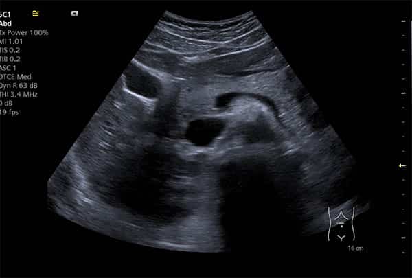
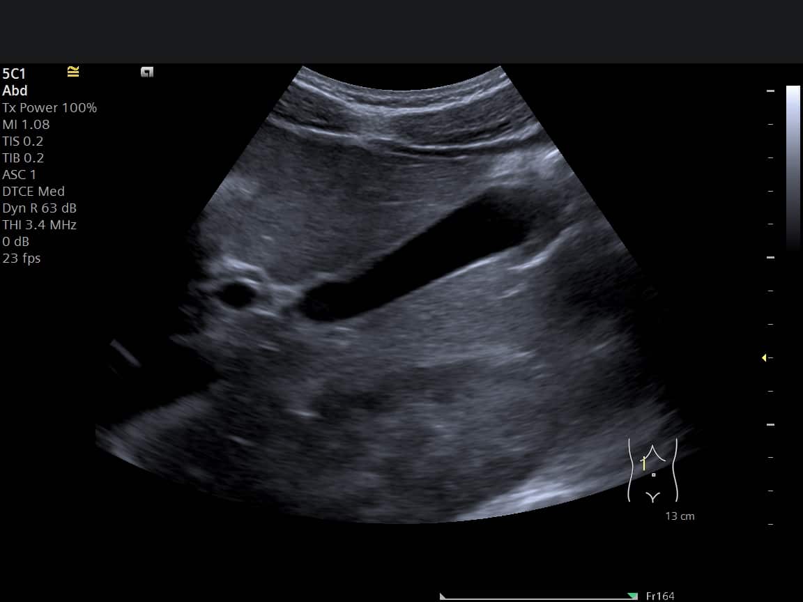
Normal reports are strikingly formulaic — and knowing the formula turns an intimidating page into good news you can recognise line by line. A typical normal whole-abdomen report reads something like: liver normal in size with smooth, homogeneous echotexture and no focal lesion; gallbladder thin-walled with no calculi (no stones); common bile duct (CBD) not dilated; pancreas partially visualised, appears unremarkable; spleen normal in size; kidneys normal in size and appearance with no hydronephrosis or calculi; aorta normal in calibre; no free fluid. Every one of those clauses is a specific check passed — the boring report is the one you want.
“Normal versus abnormal” is therefore less about single scary words and more about departures from this template — a measurement outside range, a described lesion, a “dilated” where “not dilated” belongs. The impression section at the end states whether any departure actually matters.
Echotexture — homogeneous, heterogeneous, coarse
Echotexture means the textural appearance of an organ’s tissue on screen — how evenly it reflects sound. A healthy liver looks homogeneous: smooth and uniform, like fine, even grain. Heterogeneous echotexture means the texture looks mixed or uneven; coarse echotexture means the grain looks rougher than normal. Neither word is a diagnosis — they are descriptions that prompt the question why.
Does a heterogeneous liver mean cancer? Usually not. By far the commonest associations are fatty change and inflammation, with longstanding liver conditions producing coarser textures over time; the report reads the texture alongside liver size, blood tests and your history, and says whether follow-up imaging is warranted. Whether a coarse texture can improve depends entirely on its cause — fatty change, for instance, often responds to addressing what drove it, which is a conversation for your GP with the report in hand. The full picture — causes, follow-up, and when texture findings matter — is in our dedicated guide to liver echotexture explained.
The common findings, translated
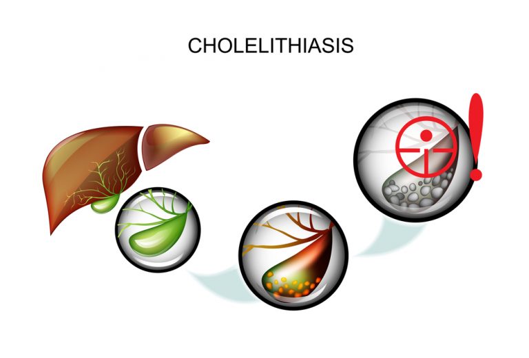
“Cholelithiasis” — gallstones, with size and number usually noted; what they look like and what happens next is covered in gallstones on ultrasound. “Fatty infiltration” / “hepatic steatosis” — fat within the liver, extremely common, graded and managed through your GP. “Simple cyst” — a thin-walled fluid sac, benign, usually needing nothing beyond being recorded. “Haemangioma” — the commonest benign liver lesion, a knot of blood vessels with a typical bright appearance. “Prominent” — radiology’s softest word: at the upper end of normal, worth recording, rarely worth worrying about. And measurements everywhere — recorded not as alarms but as baselines the next scan can be compared against.
“Partially visualised” and the limits language
“Pancreas partially visualised” is probably the most-Googled hedge in abdominal reporting — and it is normal. The pancreas sits behind the stomach and bowel, and gas blocks sound, so most scans see the head of the pancreas well and the tail incompletely. The phrase is honesty, not evasion: the report is telling your clinician exactly how far the examination could see. The same family includes “obscured by bowel gas”, “suboptimal views” (often an unfasted gallbladder or a not-full bladder — preparation matters), and “cannot exclude” — the report declining to claim certainty ultrasound cannot deliver, and naming where CT, MRI or endoscopy would be definitive if the clinical picture demands it.
Reports that admit their limits are the trustworthy ones — and if symptoms persist despite a reassuring scan, that limits language is precisely what tells your GP which test comes next. Our results guide covers taking a report forward.
Abdominal reports — your questions answered
What does heterogeneous echotexture of the liver mean?
Does a heterogeneous or coarse liver mean cancer?
What does “pancreas partially visualised” mean — should I worry?
What is actually checked in a whole-abdomen report?
My report is normal but I still have symptoms — what now?
Who explains my report to me?
The report cluster
Heterogeneous, coarse, fatty — the causes, the follow-up, and when texture matters.
Read the guide →
Start HereUnderstanding Your ReportHow every ultrasound report is structured, and the stock phrases translated.
Read the guide →
£159Abdominal Ultrasound ScanThe examination behind the report — what it covers and how to prepare.
About the scan →
A report explained before you ever read it
Findings walked through on screen at your appointment, and a written report — usually within two hours — phrased for your GP to act on. Four-hour fast, thirty minutes, done.
Kensington, London W8 4ED
3 mins from Notting Hill Gate
This guide is general information, not individual medical advice — your own report should always be interpreted with the clinician it was written for. If a report raises questions, your GP is the right first call; if in doubt about a symptom, call NHS 111.
Written and clinically reviewed by the HCPC-registered sonographers who perform this scan at IUS London — a CQC-registered diagnostic ultrasound clinic (Provider ID 1-2775844974). Your own findings are explained to you at the scan and set out in your report.
Author: Yianni Kiromitis, Senior Sonographer, HCPC RA38415
Medically reviewed: 31 July 2026