Ultrasound Glossary — Report Terms in Plain English
Every recurring term from ultrasound reports and appointments, defined honestly and without jargon — from anechoic to within normal limits.
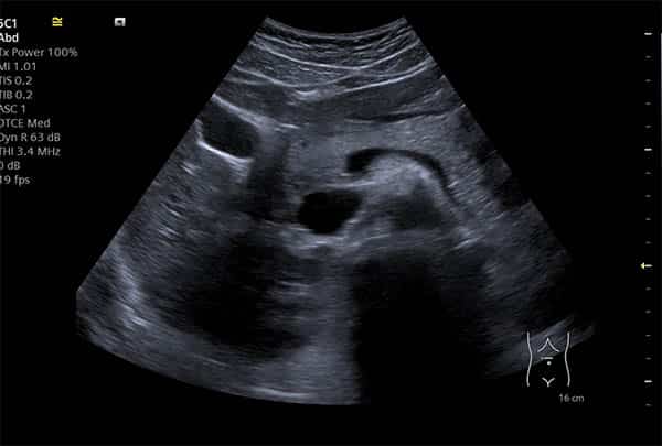
Ultrasound reports describe images in a compact technical vocabulary — precise for clinicians, opaque for everyone else. This glossary defines the terms you are most likely to meet, grouped by what they describe: how the image is made, the brightness words, the findings words, and the measurements and stock phrases reports are built from. For how a report fits together as a whole, start with our guide to understanding your ultrasound report.
How the image is made
Ultrasound (sonography). Imaging that builds live pictures from high-frequency sound echoes — no radiation involved.
Transducer (probe). The handheld device that sends sound pulses into the body and listens for the echoes that become the image.
B-mode. Standard two-dimensional grey-scale imaging — the familiar ultrasound picture.
M-mode. A mode that traces movement over time, classically used for heart motion.
Doppler. A technique that measures blood flow from frequency changes in echoes off moving blood cells.
Colour Doppler. Doppler displayed as colour overlay showing flow direction and speed. Colour means blood flow — it is not, by itself, good or bad news.
Waveform. The graph of blood flow over time produced in Doppler studies, from which flow velocities are measured.
Resolution. The machine’s ability to distinguish fine detail; higher-frequency probes see finer detail at shallower depths.
Transabdominal. Scanning through the skin of the abdomen — the external approach.
Transvaginal (TV). An internal pelvic scan using a slim probe, giving much closer, clearer views of the uterus and ovaries; explained fully in our first-time guide.
Sonographer. The healthcare professional trained to perform and interpret ultrasound examinations — in the UK, registered with the HCPC, whose public register lets you verify any sonographer’s credentials.
Thermal index (TI) & mechanical index (MI). Live safety readouts on every machine, tracking potential tissue warming and mechanical effects; kept low under the ALARA principle described in Is Ultrasound Safe?
The brightness words
Anechoic. Appears black — no echoes returned — which usually means simple fluid: a cyst, urine, a blood vessel. In context, often the most reassuring word on the page.
Hypoechoic. Darker than the surrounding tissue — fewer echoes returned. A description of brightness, not a diagnosis.
Hyperechoic / echogenic. Brighter than surroundings — strong echoes, typical of denser tissue, fat, calcification or gas.
Isoechoic. The same brightness as neighbouring tissue — sometimes noted precisely because it makes a structure subtle.
Homogeneous. Uniform in texture throughout — generally what healthy organ tissue looks like.
Heterogeneous. Mixed or uneven texture. Sometimes meaningful, often not — what it implies for the liver specifically is covered in our echotexture guide.
Echotexture. The overall textural appearance of an organ’s tissue on screen — the backdrop against which findings stand out.
Parenchyma. The working tissue of an organ (liver parenchyma, kidney parenchyma), as opposed to its coverings and plumbing.
Acoustic shadowing. The dark stripe behind something sound cannot pass through — classically a gallstone or kidney stone. Often the clue that confirms a stone.
Acoustic (posterior) enhancement. Extra brightness behind fluid, because fluid barely absorbs sound — a supporting clue that a structure is a simple cyst.
Artefact. Any image feature produced by the physics of scanning rather than by real anatomy — recognised and discounted by the sonographer.
The findings words
Cyst (simple). A thin-walled sac of fluid — anechoic, with posterior enhancement and no internal blood flow. Extremely common and benign in most locations.
Complex cyst. A cyst containing echoes, septations or solid areas — not automatically sinister, but described carefully because its features decide follow-up.
Septation. A thin internal wall dividing a cyst into compartments — one of the features weighed when grading a cyst.
Nodule. A small, discrete lump of tissue — the standard word for thyroid findings, where the overwhelming majority are benign.
Lesion. Radiology’s deliberately neutral catch-all for “an area that differs from normal tissue” — it carries no verdict at all.
Mass. A space-occupying finding, solid or mixed. Descriptive, not diagnostic — benign masses vastly outnumber sinister ones.
Calcification. Calcium deposits appearing bright, often with shadowing — common in ageing tissue, sometimes a grading feature in nodules.
Vascularity. How much blood flow a structure shows on Doppler — one feature among several, never a verdict alone.
Free fluid. Fluid outside organs. A small amount can be physiological (normal), particularly in the pelvis; larger amounts are described and investigated.
Lymphadenopathy. Enlarged lymph nodes — most commonly reactive (responding to infection), with appearance and architecture guiding whether more is needed.
Sludge. Thickened bile layering in the gallbladder — can cause gallstone-like symptoms and precede stones; covered in gallstones on ultrasound.
Polyp. A small growth from a lining surface (gallbladder, endometrium) — measured, characterised and usually simply monitored.
Hydronephrosis. Swelling of a kidney’s drainage system because urine is backing up — a sign that prompts a search for the cause of obstruction.
Varicosity. An enlarged, incompetent vein — mapped with Doppler in venous studies.
Effusion. Fluid collecting in a joint or body space — a finding that explains swelling and guides treatment.
Biopsy (ultrasound-guided). Taking a tissue sample with the needle steered live on screen — how an imaging suspicion becomes an actual diagnosis.
Measurements and report phrases
Post-void residual. Urine left in the bladder after emptying — a key number in urinary assessments, explained in our urologic guide.
Endometrial thickness. The measured depth of the womb lining, read against menstrual cycle stage or menopausal status — context is everything with this number.
CBD (common bile duct). The main bile drainage tube; its calibre is measured on abdominal scans, with dilation prompting a search for obstruction.
CRL (crown–rump length). The head-to-bottom measurement of an early pregnancy, used for dating.
EFW (estimated fetal weight). A calculated estimate from several fetal measurements in later pregnancy — an estimate with a recognised margin, tracked over time rather than read alone.
“Unremarkable”. Nothing abnormal to remark on — good news in radiology’s native understatement.
“Grossly normal”. Normal on overall inspection (“grossly” = “as a whole”, not “very”).
“Within normal limits”. Measured, and the measurement falls in the normal range.
“Cannot exclude…”. Honest acknowledgement of ultrasound’s limits — flagging where only another test could be definitive, which in context is often not needed at all.
“Clinical correlation advised”. An instruction to the receiving clinician to read the finding alongside your symptoms — routine phrasing, not a coded warning.
Incidental finding. Something unrelated to the reason for the scan — very common, mostly benign, named and sized so nothing is left vague.
“Follow-up recommended”. The finding warrants a recheck after a stated interval — surveillance, not alarm; the interval itself is the message about urgency.
Using this glossary
Is “echogenic” or “hypoechoic” bad news?
My report uses a term that isn’t here — what should I do?
Why do reports describe things instead of just giving a diagnosis?
The report guides
How every report is structured, and what the stock phrases really mean.
Read the guide →
AbdominalYour Abdominal ReportLiver, gallbladder, kidneys and pancreas phrases, translated.
Read the guide →
PelvicYour Pelvic ReportEndometrial thickness, ovaries, follicles and fibroids decoded.
Read the guide →
Reports you don’t need a glossary for
Findings explained in plain English at your appointment, and a written report — usually within two hours — phrased for your GP to act on.
Kensington, London W8 4ED
3 mins from Notting Hill Gate
Definitions are written for general patient understanding, not as clinical reference. Your own report should always be interpreted with the clinician it was written for — if in doubt, speak to your GP or call NHS 111.
Written and clinically reviewed by the HCPC-registered sonographers who perform this scan at IUS London — a CQC-registered diagnostic ultrasound clinic (Provider ID 1-2775844974). Your own findings are explained to you at the scan and set out in your report.
Author: Yianni Kiromitis, Senior Sonographer, HCPC RA38415
Medically reviewed: 31 July 2026