Kidney Transplant Imaging — What Follow-Up Scans Look For
Ultrasound is the workhorse of transplant follow-up. Here is what it assesses at each stage — and why your transplant unit stays in charge of the schedule.
Contact your transplant unit first — not a private clinic
A sudden drop in urine output, pain or swelling over the transplant, fever, or a rising creatinine on your blood tests needs your transplant team the same day. They have your history, your immunosuppression details and direct access to biopsy. Private imaging is a supplement to that relationship and never a substitute for it.

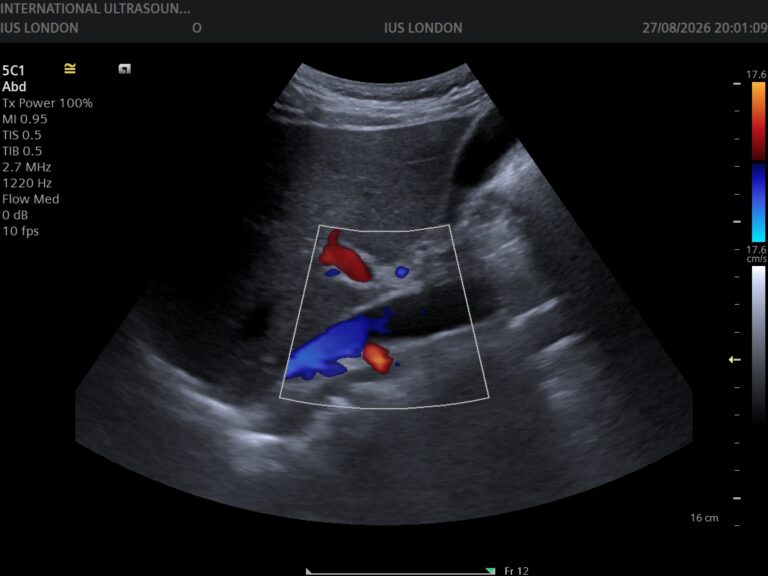
A transplanted kidney sits low in the pelvis, close to the surface — considerably easier to image than your own kidneys. That accessibility, combined with no radiation and no contrast, is why ultrasound with Doppler is the first-line test throughout transplant follow-up.
What the scan looks for, and when
| Stage | Main questions |
|---|---|
| First days after transplant | Is blood flowing into and out of the kidney? Any collection around it? Any obstruction to drainage? |
| Early weeks | Fluid collections — urinoma, haematoma, lymphocele — and whether any is pressing on the kidney or ureter |
| Any time function drops | Obstruction, collections, and the resistive index as a marker of what is happening within the kidney |
| Months to years | Narrowing of the transplant artery, changes in size and texture, stones, and the bladder |
| Long term | Routine surveillance as set by your unit, plus assessment of your own native kidneys where relevant |
What ultrasound cannot settle
The most important limitation: ultrasound cannot diagnose rejection. It can show findings that raise the question — a raised resistive index, a swollen kidney — but those appearances are non-specific and occur in several other conditions. Rejection is diagnosed on biopsy, performed by your transplant unit. Anyone implying a scan can rule rejection in or out is overstating what the test does.
Similarly, it cannot measure kidney function. That is creatinine and eGFR on blood tests, monitored by your unit. Imaging and biochemistry answer different questions, and transplant care depends on both moving together.
Where ultrasound is genuinely decisive: excluding obstruction, finding and sizing collections, and assessing blood flow. Those are the mechanical problems that need identifying quickly, and it identifies them well.
Where a private scan fits
- 1
Alongside your unit, not insteadWe are useful for interval reassurance or for a scan between hospital appointments — with your unit informed. Anything acute goes to them first.
- 2
Bring your historyTransplant date, recent creatinine, and previous scan reports. Transplant imaging is interpreted against your own baseline, and a standalone scan is much weaker without it.
- 3
The scanGreyscale and Doppler assessment of the transplant kidney low in the pelvis, plus the bladder. 30 minutes, comfortably full bladder helpful.
- 4
What we assessSize and texture, collecting system for obstruction, any fluid collection measured, arterial and venous flow with resistive index, and the bladder before and after emptying.
- 5
ResultsExplained on screen as we scan, written report usually within two hours — written for your transplant team, with values presented for direct comparison against your previous imaging.
Your questions answered
Can an ultrasound detect rejection?
What is the resistive index?
What is a lymphocele?
How often should a transplant kidney be scanned?
Does the scan check my own kidneys too?
Do I need a full bladder?
Where to go from here
Doppler assessment, reported for your unit
Transplant kidney size and texture, obstruction, collections and full Doppler flow assessment with resistive index — explained on screen as we scan, with the report usually within two hours in a form your transplant team can use.
Kensington, London W8 4ED
3 mins from Notting Hill Gate
This article is general information about transplant kidney imaging, not individual medical advice. Any drop in urine output, pain over the transplant, fever or rising creatinine needs your transplant unit the same day. Ultrasound cannot diagnose rejection — that requires biopsy. See NHS guidance on kidney transplant. We do not offer biopsy, blood tests or CT.
Written and clinically reviewed by the HCPC-registered sonographers who perform this scan at IUS London — a CQC-registered diagnostic ultrasound clinic (Provider ID 1-2775844974). Your own findings are explained to you at the scan and set out in your report.
Author: Yianni Kiromitis, Senior Sonographer, HCPC RA38415 — over 20 years’ experience in NHS and private ultrasound
Medically reviewed: 9 August 2026