Armpit Lump — Lymph Node, Cyst or Lipoma?
Most armpit lumps are lymph nodes doing their job. Here is what the different kinds feel like, when to wait, and when to stop waiting.
The armpit contains a cluster of lymph nodes that drain the arm, the chest wall and the breast. They swell when the immune system is busy — a cut on the hand, a skin infection, a viral illness, sometimes a recent vaccination in that arm. That accounts for the majority of armpit lumps, and most settle within a few weeks.
The armpit also holds sweat glands, hair follicles and fat, so cysts, blocked glands and lipomas turn up here too. Each has a different feel and a different story.
What the different lumps feel like
| What you notice | Commonly |
|---|---|
| Tender, appeared with an illness, cut or vaccination; shrinking over a few weeks | Reactive lymph node |
| Soft, doughy, moves easily under the skin, painless, slow-growing over years | Lipoma — a benign fatty lump |
| Attached to the skin, sometimes with a small central dot; may become red and sore | Sebaceous or epidermoid cyst |
| Recurring painful boils and tunnels in the armpit or groin | Hidradenitis suppurativa — see your GP, it is treatable |
| Firm, painless, fixed, persisting or growing over weeks | Needs assessment — see your GP |
| Soft swelling more noticeable before periods, sometimes tender | Accessory breast tissue |
See your GP if it lasts more than three weeks
Go sooner if the lump is hard, fixed or growing, if there is also a breast change, or if you have unexplained weight loss, night sweats or persistent fever. Armpit nodes drain the breast, so a new armpit lump alongside any breast change should be assessed together on the NHS breast clinic pathway. A hot, spreading, painful swelling with fever needs same-day care.
What ultrasound settles
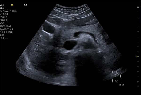
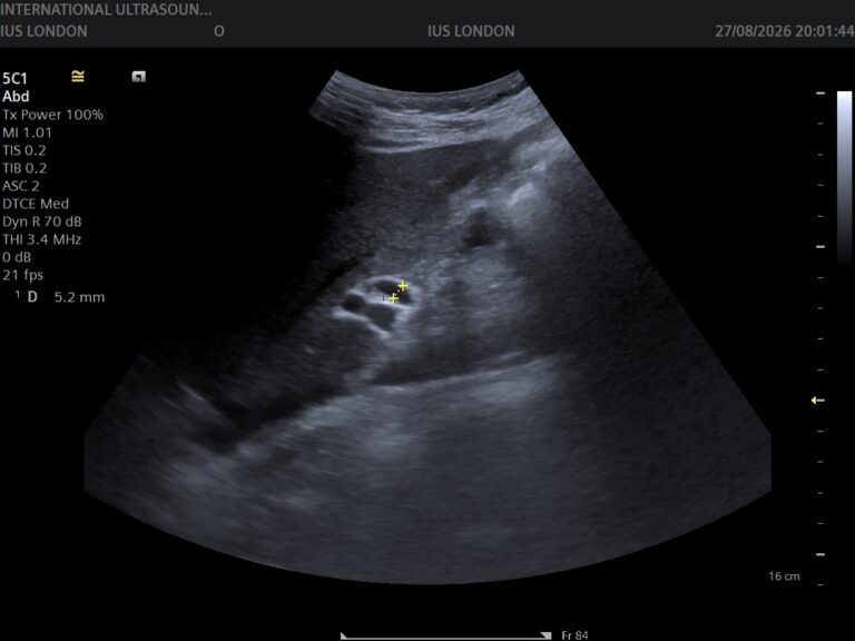
Everything in the armpit sits close to the surface, which suits ultrasound well. A scan establishes which structure the lump is — node, cyst, lipoma or breast tissue — and measures it. For lymph nodes it assesses the features that matter: size, shape, whether the normal internal architecture is preserved, and the pattern of blood flow. Reactive nodes and suspicious nodes look meaningfully different.
What a scan cannot do is give a tissue diagnosis. Where a node looks abnormal, sampling is the next step — done at a breast clinic or by an appropriate specialist, not here. We do not perform biopsy or fine needle aspiration, and a reassuring-looking node on a scan does not override a clinician’s concern on examination.
How the appointment runs
- 1
BookingNo referral, no preparation, 30 minutes. Tell us where the lump is and how long it has been there.
- 2
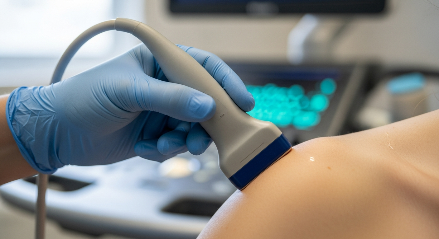
The scanLying back with the arm raised. Warm gel and a small probe over the armpit — painless, and we work gently around anything tender.
- 3
What we assessThe lump identified, measured and characterised, the other nodes in the armpit, and the adjacent breast tissue where relevant. The other side is often scanned for comparison.
- 4
ResultsExplained on screen as we scan, with the written report usually with you within two hours — and if a node needs sampling, the report says so plainly for your GP.
Your questions answered
How long should I wait before getting an armpit lump checked?
Can a vaccination cause an armpit lump?
Should I worry if it is painless?
Could it be related to my breast?
What is a lipoma?
Do you remove lumps?
Where to go from here
Know what it is
The lump identified, measured and characterised alongside the surrounding nodes and tissue in a 30-minute appointment — explained on screen as we scan, with the report usually within two hours for your GP.
Kensington, London W8 4ED
3 mins from Notting Hill Gate
This article is general information about armpit lumps, not individual medical advice. A lump lasting more than three weeks, or one that is hard, fixed or growing, should be assessed by your GP — see NHS guidance on swollen glands and breast cancer. We do not offer biopsy, fine needle aspiration or removal of lumps.
Written and clinically reviewed by the HCPC-registered sonographers who perform this scan at IUS London — a CQC-registered diagnostic ultrasound clinic (Provider ID 1-2775844974). Your own findings are explained to you at the scan and set out in your report.
Author: Yianni Kiromitis, Senior Sonographer, HCPC RA38415 — over 20 years’ experience in NHS and private ultrasound
Medically reviewed: 9 August 2026