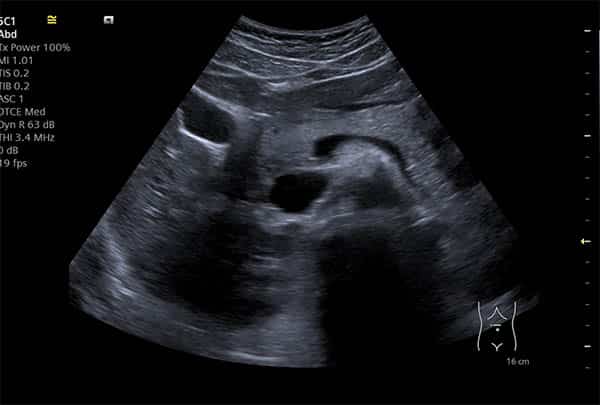
Raised CA-125 — Why a Pelvic Ultrasound Comes First
Book a Private Scan
Same-day appointments · Kensington, London · No GP referral needed
Private Pelvic Ultrasound ScanUnderstanding Raised CA-125 Levels: Your First Steps
Receiving news of a raised CA-125 blood test result can be concerning. It's natural to feel anxious and wonder what this means for your health. CA-125 (Cancer Antigen 125) is a protein found in the blood that can be elevated in various conditions, both benign and malignant. While it's often associated with ovarian cancer, it's crucial to understand that a raised CA-125 level alone does not confirm a cancer diagnosis.
This article will guide you through understanding CA-125, why a pelvic ultrasound is typically the first and most important next step, and how IUS London supports you in this journey. Our aim is to provide clear, patient-centred information to help you navigate this situation with confidence.
Red-Flag / Urgent Care Information
If you are experiencing any sudden, severe, or worsening symptoms such as acute pelvic pain, abdominal swelling, persistent indigestion, or unexplained weight loss, please seek urgent medical attention from your GP or an emergency department immediately. While this article discusses investigations for raised CA-125, acute symptoms require prompt clinical assessment.
What is CA-125 and What Does a Raised Level Mean?
CA-125 is a glycoprotein produced by certain cells in the body. It is present in small amounts in the blood of most healthy women. However, its levels can become elevated in response to inflammation, irritation, or the presence of certain conditions, including some cancers.
It's important to recognise that CA-125 is non-specific. This means that while it can be a marker for ovarian cancer, many other conditions can also cause its levels to rise. This non-specificity is why further investigation, particularly a pelvic ultrasound, is so vital.
Common Benign Causes of Raised CA-125
Many non-cancerous conditions can lead to an elevated CA-125. These include:
- Menstruation: Levels can fluctuate during the menstrual cycle.
- Pregnancy: Especially during the first trimester.
- Endometriosis: A condition where tissue similar to the lining of the womb grows outside the uterus.
- Uterine fibroids: Non-cancerous growths in the uterus.
- Pelvic inflammatory disease (PID): An infection of the female reproductive organs.
- Liver disease: Conditions like cirrhosis or hepatitis.
- Pancreatitis: Inflammation of the pancreas.
- Diverticulitis: Inflammation or infection of pouches in the digestive tract.
- Peritonitis: Inflammation of the lining of the abdomen.
- Ovarian cysts: Most ovarian cysts are benign and can cause CA-125 elevation.
Symptoms Associated with Ovarian Concerns
While a raised CA-125 can be asymptomatic, some women may experience symptoms that prompt the blood test. These can include:
- Persistent abdominal bloating
- Feeling full quickly or difficulty eating
- Pelvic or abdominal pain
- Changes in bowel habits (e.g., constipation or diarrhoea)
- Increased urinary frequency or urgency
- Unexplained weight loss or gain
- Fatigue
It's crucial to discuss any persistent or new symptoms with your doctor, regardless of your CA-125 levels.
The Crucial Role of Pelvic Ultrasound
When a CA-125 level is found to be elevated, a pelvic ultrasound scan is almost always the next recommended step. This imaging technique uses sound waves to create detailed pictures of the organs within your pelvis, including the uterus, fallopian tubes, and ovaries. It is a non-invasive, safe, and highly effective way to visualise these structures.
Why Ultrasound Comes First
- Visualisation of Ovaries and Pelvic Organs: Ultrasound provides immediate visual information about the size, shape, and structure of the ovaries. It can detect cysts, masses, or other abnormalities that might explain the raised CA-125.
- Distinguishing Benign from Malignant: Experienced sonographers and radiologists can often differentiate between benign (non-cancerous) and suspicious (potentially cancerous) ovarian masses based on specific ultrasound features. Features like solid components, septations, and blood flow patterns are assessed.
- Risk of Malignancy Index (RMI): The RMI is a scoring system used to assess the risk of an ovarian mass being malignant. It combines three factors: the CA-125 level, menopausal status, and ultrasound findings. A higher RMI score indicates a greater likelihood of malignancy, guiding further management decisions. The ultrasound component is critical for this calculation.
- Guiding Further Management: The ultrasound findings directly influence the next steps. If the ultrasound shows clear benign features, it can provide significant reassurance and may avoid unnecessary invasive procedures. If suspicious features are noted, it helps in planning appropriate referral to a gynaecological oncologist.
Management, Context, and Pathways Following a Pelvic Ultrasound
Once a pelvic ultrasound has been performed, the findings, in conjunction with your CA-125 level and clinical picture, will guide the next steps in your management. The pathway chosen will depend on whether the ultrasound indicates benign features, suspicious features, or is inconclusive.
Interpreting Ultrasound Findings
Benign Features: If the ultrasound clearly shows features consistent with a benign condition (e.g., a simple ovarian cyst, fibroids), and your CA-125 elevation is mild, your doctor may recommend a period of watchful waiting, repeat imaging, or conservative management. Many benign ovarian cysts resolve on their own.
Suspicious Features: If the ultrasound reveals features that are concerning for malignancy (e.g., solid components, abnormal blood flow, ascites), or if the RMI score is high, you will likely be referred to a gynaecological oncologist for further evaluation. This may involve additional imaging (such as an MRI or CT scan) and potentially surgical intervention.
Inconclusive Findings: Sometimes, the ultrasound may not provide a definitive answer. In such cases, further investigation, including advanced imaging or specialist consultation, may be necessary to clarify the diagnosis.
The Importance of a Multidisciplinary Approach
Managing a raised CA-125 and suspicious pelvic findings often involves a multidisciplinary team. This team may include:
- General Practitioner (GP): Your primary care physician who initiates investigations and coordinates care.
- Gynaecologist: A specialist in female reproductive health.
- Radiologist/Sonographer: Experts in medical imaging who perform and interpret scans.
- Gynaecological Oncologist: A surgeon specialising in cancers of the female reproductive system.
- Pathologist: A doctor who examines tissue samples to make a definitive diagnosis.
This collaborative approach ensures that you receive comprehensive and individualised care, leading to the most accurate diagnosis and appropriate treatment plan.
IUS London's Role: From Our Practice
At IUS London, our private ultrasound clinic in Kensington is dedicated to providing rapid, accurate, and reassuring diagnostic services. When faced with a raised CA-125, our priority is to offer a prompt and thorough pelvic ultrasound, often available on the same day or usually within two hours, to alleviate anxiety and guide immediate next steps.
Our experienced sonographers utilise modern ultrasound technology to meticulously assess your pelvic organs. We focus on detailed imaging of the ovaries, uterus, and surrounding structures, looking for any features that might explain the CA-125 elevation. Our reports are comprehensive and include all necessary measurements and descriptions to inform your referring clinician.
Experiential Claim: Anonymised Presentation Pattern
We recently saw a 48-year-old pre-menopausal patient referred with a CA-125 of 85 U/mL. Clinically, she reported mild, intermittent pelvic discomfort. Our pelvic ultrasound revealed a 5cm unilocular ovarian cyst with no solid components or abnormal vascularity. Given the benign ultrasound appearance and her pre-menopausal status, a diagnosis of a functional ovarian cyst was made, and a follow-up scan was recommended. This rapid assessment provided significant reassurance and avoided unnecessary invasive procedures.
Comparison of Diagnostic Tools for Ovarian Assessment
To further illustrate why ultrasound is the primary investigation, consider the strengths and limitations of various diagnostic tools:
| Diagnostic Tool | Strengths | Limitations | Role in Pathway |
| **CA-125 Blood Test** | Easily accessible, quantitative marker | Non-specific, elevated in many benign conditions, can be normal in early cancer | Initial screening, part of RMI, monitoring |
| **Pelvic Ultrasound** | Non-invasive, real-time imaging, detailed anatomical view | Operator-dependent, limited by patient body habitus, cannot definitively diagnose malignancy | First-line imaging, crucial for RMI, guides further investigation |
| **MRI Scan** | Excellent soft tissue contrast, no radiation | More expensive, longer scan time, not always readily available | Second-line imaging for complex cases, surgical planning |
| **CT Scan** | Good for assessing spread of disease, rapid | Involves ionising radiation, less detailed for ovarian morphology | Assessing disease extent, staging, not primary ovarian assessment |
| **Biopsy/Surgery** | Definitive diagnosis | Invasive, carries risks, usually performed after imaging suggests malignancy | Confirmatory diagnosis, treatment |
Call to Action (CTA) Section
If you have received a raised CA-125 result, or if you are experiencing pelvic symptoms, don't delay seeking further investigation. A timely and accurate pelvic ultrasound is crucial for understanding your health and guiding appropriate management.
- Book Your Pelvic Ultrasound: Schedule your private pelvic ultrasound scan at IUS London today. Private Pelvic Ultrasound Scan
- Learn More About Our Services: Explore our full range of women's health scans. Women's Scans
- Contact Us: Our friendly team is here to answer your questions and provide support. Contact IUS London
References
- van Gorp, T., et al. (2012). Subjective assessment by ultrasound is superior to the risk of malignancy index (RMI) or the risk of ovarian malignancy algorithm (ROMA) in discriminating benign from malignant adnexal masses. European Journal of Cancer, 48(11), 1649-1656. https://consensus.app/papers/details/b2611485ee0e56d88ca49bb0d87ea7d2/
- Jacobs, I. J., et al. (1990). A risk of malignancy index incorporating CA 125, ultrasound and menopausal status for the accurate preoperative diagnosis of ovarian cancer. BJOG: An International Journal of Obstetrics & Gynaecology, 97(10), 922-929. https://consensus.app/papers/details/fcacf693bbc05dab8593f2d8f1f6c30c/
- Maggino, T., et al. (1994). Prospective multicenter study on CA 125 in postmenopausal pelvic masses. Gynecologic Oncology, 54(2), 117-122. https://consensus.app/papers/details/7c3dd174bf1c5036a1ce18e19bd6adbb/
- Buys, S. S., et al. (2011). Effect of screening on ovarian cancer mortality: the Prostate, Lung, Colorectal and Ovarian (PLCO) Cancer Screening Randomized Controlled Trial. JAMA, 305(22), 2295-2303. https://consensus.app/papers/details/0d70216bdace5dabad778a59d3b43d84/