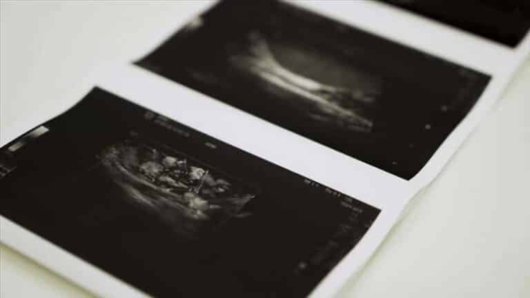
Uterine Fibroids on Ultrasound — What a Scan Shows
Ultrasound is the first-line test for fibroids. What it sees, what it can miss, where fibroids sit and why location matters more than size — plus straight answers to the questions people are embarrassed to ask.
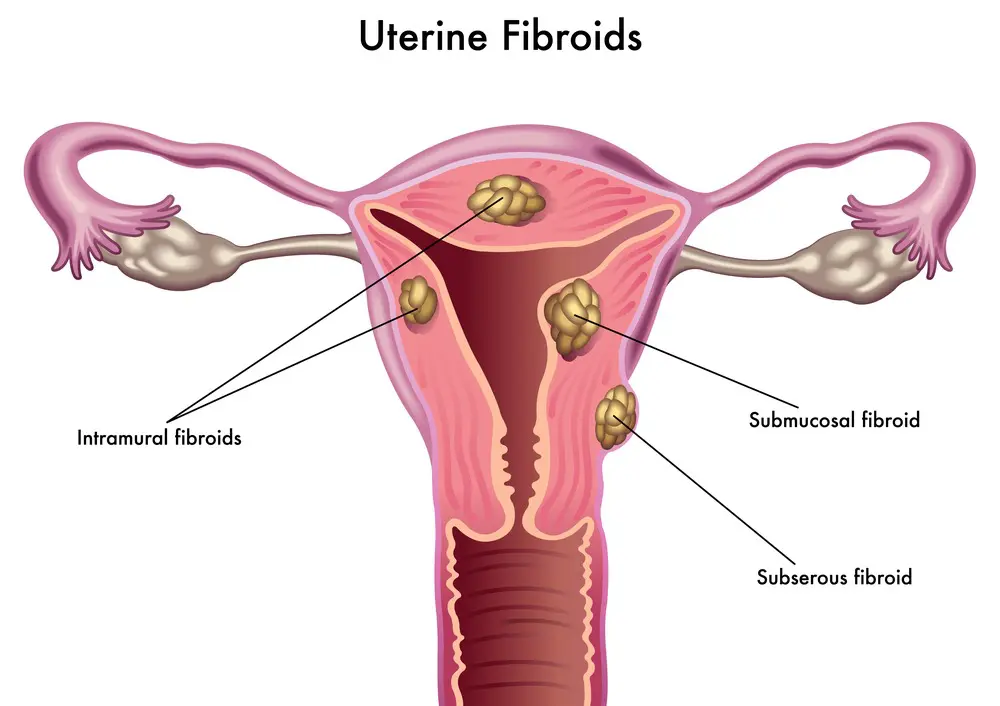
Yes — fibroids show up on ultrasound, and ultrasound is the standard first-line test for diagnosing them. They have a characteristic appearance: well-defined, rounded areas of muscle tissue, usually slightly darker than the surrounding womb wall, often with a distinctive pattern of shadowing. A scan can confirm they are present, count them, measure each one, and — most usefully — establish exactly where each sits, which determines whether they explain your symptoms at all.
Fibroids are extremely common, benign growths of the muscular wall of the womb. Many women have them without ever knowing. The purpose of imaging is not simply to find them, but to work out whether the ones you have account for what you are experiencing.
The questions people hesitate to ask
Are fibroids sexually transmitted? No. Fibroids are not an infection and cannot be passed between people in any way. They are growths of your own uterine muscle, driven largely by hormones and genetics. Chlamydia and other sexually transmitted infections do not cause fibroids — that is a completely separate condition affecting different tissue, covered in our guide to pelvic infection. Nothing about having fibroids says anything about your sexual history.
Are fibroids cancerous? Almost never. Fibroids are benign, and having them does not raise your risk of developing cancer of the womb. A malignant muscle tumour of the uterus does exist but is genuinely rare, and rapid growth — particularly after menopause — is the feature that prompts closer assessment. If a scan raises that question, you will be referred rather than watched.
Did I cause them? No. Fibroids are not caused by diet, stress, contraception, or anything you did or failed to do. Risk is influenced by age, family history, ethnicity and hormonal factors — none of which is a choice.
Where a fibroid sits matters more than how big it is
This is the single most useful thing a scan report tells you, and the part most often glossed over. A small fibroid in the wrong place causes far more trouble than a large one sitting harmlessly on the outside.
| Type | Where it sits | What it typically causes |
|---|---|---|
| Submucosal | Bulging into the cavity of the womb | The most symptomatic type — heavy bleeding, and the type most associated with fertility and implantation problems, even when small |
| Intramural | Within the muscular wall | Heavy or painful periods and pressure symptoms once sizeable |
| Subserosal | On the outer surface | Often silent; may press on bladder or bowel when large |
| Pedunculated | On a stalk, inside or outside | Can cause sudden pain if it twists on its stalk |
| Cervical | In the neck of the womb | Uncommon; may cause discharge, bleeding or discomfort |
A report describing several fibroids will usually locate and measure each. Reading it alongside our guide to pelvic reports makes the terminology considerably less opaque — including phrases like “bulky uterus”, which usually just means fibroids have enlarged it.
Can fibroids be missed on a scan?
Yes, occasionally — and since people search for exactly this, here is the honest picture rather than a reassuring one.
Very small fibroids may not be resolved, particularly a few millimetres across. A womb already enlarged by multiple fibroids can be difficult to assess completely, because the larger ones obscure what lies behind them — and counting becomes approximate. Scanning route matters: a transvaginal scan sees the cavity and smaller fibroids far better than scanning through the abdomen, while a very large fibroid uterus may actually need the abdominal view to capture its full extent. Both are often used together.
Submucosal fibroids are the ones where certainty matters most, because they drive bleeding and fertility problems — and they are also the ones where a standard scan can under-call the extent to which a fibroid indents the cavity. Where that distinction changes management, a saline infusion scan, hysteroscopy or MRI may be arranged through a gynaecologist. MRI is also used for surgical planning, where precise mapping is needed.
If your symptoms and your scan do not match — heavy bleeding with a scan reported as normal, for instance — that mismatch is worth raising with your GP rather than accepting. Other conditions, including other causes of heavy periods, may be responsible.
What fibroids feel like — when they feel like anything
Most fibroids cause no symptoms whatsoever and are found incidentally during a scan arranged for another reason. Where they do cause problems, the usual pattern is:
- Heavy or prolonged periods — the commonest complaint, sometimes with clots or flooding, and occasionally enough to cause iron-deficiency anaemia
- Pelvic pressure or a feeling of fullness, or a noticeably firmer lower abdomen
- Needing to pass urine frequently, from pressure on the bladder
- Pain during sex, or a persistent ache in the pelvis or lower back
- Constipation, where a fibroid presses backwards on the bowel
- Difficulty conceiving or recurrent miscarriage — principally with submucosal fibroids distorting the cavity
Fibroids typically shrink after menopause as oestrogen falls. New growth or new bleeding after menopause is different, and should always be assessed — see postmenopausal bleeding.
Timing, monitoring and treatment
When to scan. There is no strict rule, but scanning in the first half of the cycle, soon after a period, generally gives the clearest view of the cavity when the lining is thin — useful when submucosal fibroids are the question. For simply confirming and measuring fibroids, timing matters much less.
Monitoring. Many fibroids need nothing more than periodic measurement to check they are stable, using consistent technique so that comparisons mean something. Our guide to monitoring fibroids covers how that works in practice.
Treatment is decided by symptoms rather than by the presence of fibroids, and is arranged through your GP or a gynaecologist. Options range from medication controlling bleeding, through procedures such as uterine artery embolisation, to surgical removal of fibroids or of the womb. Asymptomatic fibroids generally need no treatment at all — and a scan finding them is not, by itself, a reason to do anything.
Your questions answered
Do fibroids show up on ultrasound?
Can fibroids be missed on an ultrasound?
Are uterine fibroids sexually transmitted?
Can fibroids be cancerous?
Is there a blood test for fibroids?
What is the best time in my cycle for a fibroid scan?
Do fibroids need to be removed?
Will fibroids affect my fertility?
Where to go from here
How serial measurement works, and what counts as meaningful change.
Read the guide →
SymptomCauses of Heavy PeriodsFibroids are one cause among several — what else to consider.
Read the guide →
£159Pelvic Ultrasound ScanThe examination that finds and measures fibroids.
About the scan →
Fibroids found, located and measured
Transabdominal and transvaginal scanning by HCPC-registered sonographers, each fibroid located and measured, findings explained on screen, and a written report usually within two hours for your GP.
Kensington, London W8 4ED
3 mins from Notting Hill Gate
This article is general information, not individual medical advice. Fibroid treatment decisions should be made with your GP or a gynaecologist, guided by your symptoms. New bleeding after the menopause, or rapid growth of a fibroid, should always be assessed promptly.
Written and clinically reviewed by the HCPC-registered sonographers who perform this scan at IUS London — a CQC-registered diagnostic ultrasound clinic (Provider ID 1-2775844974). Your own findings are explained to you at the scan and set out in your report.
Author: Yianni Kiromitis, Senior Sonographer, HCPC RA38415
Medically reviewed: 1 August 2026