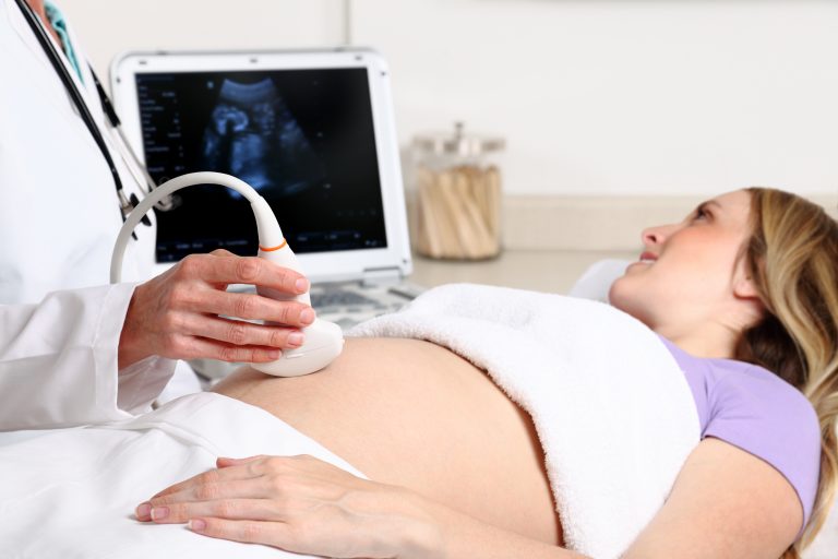
Everyone is aware these days of the use of ultrasound in medicine, although most people will associate ultrasound with pregnancy, as all pregnant women are offered at least two baby scans as part of the NHS prenatal care. The spectrum of ultrasound imaging, however, today is so wide, and it is not confined only to medicine but includes applications in engineering and other disciplines.
Most people, however, excluding sonographers and physicists, are not aware of how ultrasound was discovered and how it became such a common medical imaging procedure.
Lett take a look back in history to find out how sound waves became the diagnostic tool used in private ultrasound clinics and hospitals all over the world today.
Who discovered Ultrasound?
The first paper about ultrasound was published in 1794 by Lazzaro Spallanzani an Italian physiologist and biologist who studied bats and discovered that they use sound to navigate instead of sight. This property is called echolocation, and it is the foundation that governs today's medical ultrasound.
In 1826 a Swiss physicist name Jean Daniel Colladon used a church bell underwater and discovered that sound travels faster in the water than in the air.
Christian Andreas Doppler, an Austrian physicist and mathematician, discovered in 1842 that the frequency of a sound wave depends on the speed of the source. This principle is today known s the Doppler effect.
In 1880 Pierre Curie and his brother Jacques Curie, demonstrated the first piezoelectric effect, which is the ability of certain materials to generate an electric charge in response to mechanical stress, using crystals of tourmaline, quartz, topaz, cane sugar, and Rochelle salt.
The first working sonar system in the United States was built to detect icebergs by Reginald Fessenden in 1914. This was followed by the detection of submarines in 1917 and the detection of aircraft in 1935.
The use of ultrasound in medicine was first evaluated in 1942 by Karl Theodore Dussik to diagnose brain tumours, and in 1949 George Döring Ludwig, used ultrasound to detect gallstones.
The use of ultrasound in medicine accelerated, and its use was established in urology and cardiac applications in 1958; Ian Donald published an article in The Lancet titled Investigation of Abdominal Masses by Pulsed Ultrasound. Donald uses ultrasound to detect abdominal tumours and cysts and later is able to detect a twin pregnancy. Ian Donald becomes the father of OB-GYN ultrasound.
Ultrasound History Timeline
You can see a detailed timeline of the history of ultrasound below.
Echolocation
Lazzaro Spallanzani a physiologist, was the first to study echolocation among bats, which is the fundamental principle of ultrasound.
Piezoelectricity
Pierre and Jacques Currie discover piezoelectricity a technology used in ultrasound probes to emit and receive sound waves.
Hydrophone
Physicist Paul Langevin was commissioned to invent a device that detected objects at the bottom of the sea as a result of the sinking of the Titanic. He invented the hydrophone which today is perceived as the first transducer.
Sonography used for Therapy
Sonography was used as a treatment, to appease arthritic pain and eczema and to sterilize vaccines
First Medical Diagnosis using Ultrasound
Neurologist Karl Dussik was the first to use sonography for medical diagnoses in his attempts to detecting brain tumours.
A-Mode Ultrasound
George D. Ludwig, M.D. was the developer of A-mode ultrasound used to detect gallstones.
B-Mode Ultrasound
Douglas Howry and Joseph Holmes, were the leading pioneers of B-mode ultrasound equipment and John Reid and John Wild invented a handheld B-mode device to detect breast tumours.
First Echocardiogram
Physician Inge Edler and Engineer C. Hellmuth Hertz performed the first successful echocardiogram
Ultrasound in Ob/Gyn
Dr. Ian Donald incorporated ultrasound into the OB/GYN field of medicine.
Doppler Technique
Don Baker, Dennis Watkins, and John Reid designed pulsed Doppler ultrasound technology to image blood flow
CWD, Spectral and Colour Doppler
In the 1970s there were nmany advances of ultrasound technology including the continuous wave Doppler, spectral wave Doppler and colour Doppler ultrasound.
3D Ultrasound
Kazunori Baba developed 3D ultrasound technology and captured three-dimensional images of a foetus.
Ultrasound in Critical Care
Daniel Lichtenstein introduced lung and general sonography in intensive care units.
3D/4D
Ultrasound technology advanced improving 2D and 3D image quality. These improvements lead to the introduction of 4D ultrasound.
Mobile Sonography
Ultrasound technology never stopped improving and today ultrasound images can be obtained not only with laptop ultrasound scanners but also with wireless probes and mobile phones.