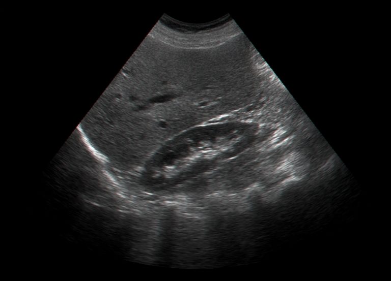
Breast Cysts — and How Ultrasound Tells Them Apart
Most breast lumps are not cancer, and cysts are among the commonest causes. What they are, how a scan distinguishes a simple cyst from something needing more, and what happens next.
Finding a lump in your breast is frightening, and no amount of reassuring statistics changes that in the moment. So the useful facts first: most breast lumps are benign, and breast cysts — fluid-filled sacs within the breast tissue — are one of the commonest causes, particularly between the ages of about 35 and 50. Ultrasound is very good at this specific question. A simple cyst has an appearance so characteristic that it can usually be identified with confidence in the same appointment, which is why a scan often ends the worry the day it starts.
Whatever you read here, a new breast lump should be assessed by your GP, who can refer you into the NHS breast clinic pathway. Go promptly rather than waiting if you notice: a hard or fixed lump that does not move · a change in breast size or shape · skin dimpling, puckering, redness or scaling · nipple inversion or discharge, particularly bloody · persistent pain unrelated to your cycle · swelling in the armpit or above the collarbone. A private scan can run alongside that referral — it should never replace it.
Breast cysts, and why they form
Breast tissue contains lobules that produce milk and ducts that carry it. A cyst forms when fluid collects in one of these structures and the surrounding tissue seals it into a sac. They are strongly influenced by hormones, which explains their pattern: most common in the years approaching menopause, often fluctuating with the menstrual cycle, frequently more tender in the week before a period, and usually settling after menopause unless you are taking HRT.
They often occur alongside fibrocystic change — generalised lumpiness and tenderness that is a normal variation rather than a disease. A cyst can appear over days, change size, and sometimes disappear on its own. Having cysts does not increase your risk of breast cancer, and having one does not mean you will develop more.
How ultrasound classifies what it finds
This is where ultrasound earns its place, because the distinction it draws is precise and immediate.
| What it is | How it looks | What usually follows |
|---|---|---|
| Simple cyst | Completely black (anechoic), thin smooth wall, brighter tissue behind it, no blood flow inside | Benign. Reassurance, and normally no further imaging needed |
| Complicated cyst | Fluid containing fine internal echoes or debris, still no solid parts | Almost always benign. Sometimes a short-interval repeat scan, or drainage if uncomfortable |
| Complex cystic and solid | Fluid with genuine solid components, thick walls or internal blood flow | Needs tissue diagnosis — referral for biopsy through your GP or breast clinic |
| Solid lump | No fluid; features assessed on margins, shape and vascularity | Many are benign (such as a fibroadenoma); assessment decides whether biopsy is needed |
The word complicated causes unnecessary alarm and is worth translating: it describes the fluid’s appearance, not the seriousness. A complicated cyst is fluid with a bit of debris in it — still overwhelmingly benign. It is complex cystic and solid lesions, with genuine solid tissue inside, that change the plan.
Aspiration, monitoring and reassurance
Most simple cysts need nothing at all. They are documented, explained, and left alone. If a cyst is painful, large, or you simply want it gone, it can be drained with a fine needle under ultrasound guidance — a procedure arranged through a breast clinic, in which the fluid is withdrawn and the lump usually disappears immediately. Cysts can refill, and that is not a sign anything is wrong.
Where the appearance is not straightforwardly benign, the honest step is tissue diagnosis rather than more scanning. Our reports state this plainly with the recommended next test, so your GP can refer without a second opinion first. Imaging describes; only a biopsy diagnoses.
Age shapes the approach too. Under 40, dense breast tissue makes ultrasound the more useful first test — covered in our guide to breast ultrasound for women under 40. From around 40 upwards, mammography and ultrasound are complementary, and breast density affects what each one shows.
What a cyst appointment usually looks like
The less common outcome is handled the same way, without euphemism: the features are described plainly, the report states that tissue diagnosis is needed, and the routing to a breast clinic happens the same day. These are generalised patterns from our practice, not individual cases; your own findings are always explained to you at your scan and set out in your report.
Your questions answered
Can ultrasound tell if a breast lump is a cyst?
Are breast cysts dangerous?
What does a “complicated cyst” mean on my report?
Do breast cysts need to be drained?
Will my cyst come back?
Should I see my GP or book a private scan?
Do I need a mammogram as well?
Where to go from here
The examination itself — what it covers and how to book.
About the scan →
Under 40Breast Ultrasound Under 40Why dense tissue makes ultrasound the better first test.
Read the guide →
ArmpitArmpit Lump — What Is It?Lymph node, cyst or something else — how the scan tells them apart.
Read the guide →
A lump answered the same appointment
Breast ultrasound by HCPC-registered sonographers, the finding shown to you on screen and explained in proportion, with a written report usually within two hours for your GP.
Kensington, London W8 4ED
3 mins from Notting Hill Gate
This article is general information, not individual medical advice. Any new breast lump or breast change should be assessed by your GP, who can refer you into the NHS breast clinic pathway; imaging describes findings, but only a biopsy provides a diagnosis. If in doubt, speak to your GP or call NHS 111.
Written and clinically reviewed by the HCPC-registered sonographers who perform this scan at IUS London — a CQC-registered diagnostic ultrasound clinic (Provider ID 1-2775844974). Your own findings are explained to you at the scan and set out in your report.
Author: Yianni Kiromitis, Senior Sonographer, HCPC RA38415
Medically reviewed: 1 August 2026