Ultrasound vs MRI vs CT — Which Scan Do I Need?
Navigating the world of medical imaging can be daunting. When your doctor recommends a scan, understanding the differences between an Ultrasound, MRI, and CT can help alleviate anxiety and empower you to ask informed questions. Each modality offers a unique window into the human body, excelling in different diagnostic scenarios. This comprehensive guide will demystify these powerful tools, helping you understand which scan might be right for your specific needs.
Understanding the Core Technologies: A Clinical Deep Dive
To truly appreciate the diagnostic power of Ultrasound, MRI, and CT, it's essential to understand the fundamental principles behind each technology and the specific anatomical or pathological insights they provide.
Ultrasound: The Real-Time, Radiation-Free Window
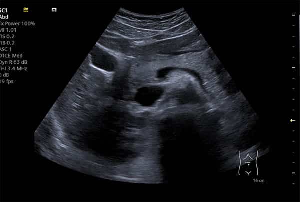
Clinical Explanation: Ultrasound, or sonography, employs high-frequency sound waves (typically 2-18 MHz) generated by a transducer. These sound waves travel into the body, encounter tissues and structures, and then echo back to the transducer. The time it takes for the echoes to return, and their intensity, are processed by a computer to create real-time images. This dynamic imaging capability is crucial for observing movement, such as blood flow within vessels (Doppler ultrasound) or the beating of a fetal heart. It's particularly adept at visualising soft tissues and fluid-filled structures, as sound waves transmit well through liquids but are reflected by bone and air. Pathologically, ultrasound is excellent for detecting cysts, tumours (differentiating solid from cystic masses), inflammation (e.g., appendicitis, cholecystitis), and assessing organ size and texture (e.g., liver, kidneys, thyroid). Its real-time nature also makes it invaluable for guiding biopsies and aspirations, ensuring precise needle placement. The absence of ionising radiation makes it a safe choice for pregnant women and children, and for repeated examinations.
Magnetic Resonance Imaging (MRI): Unparalleled Soft Tissue Contrast
Clinical Explanation: MRI is a sophisticated imaging technique that utilises a powerful magnetic field and radio waves to generate detailed images of organs and soft tissues. The body is primarily composed of water molecules, which contain hydrogen atoms. In a strong magnetic field, the protons within these hydrogen atoms align. Short bursts of radiofrequency waves are then emitted, knocking these protons out of alignment. When the radiofrequency pulse is turned off, the protons relax back into alignment, releasing energy signals. Different tissues relax at different rates, and these varying signals are detected by the MRI scanner and converted into highly detailed cross-sectional images. This technique provides superior soft tissue contrast compared to CT, making it the gold standard for visualising the brain, spinal cord, nerves, muscles, ligaments, tendons, and internal joint structures. Pathologically, MRI is exceptional for diagnosing conditions like multiple sclerosis, brain tumours, spinal disc herniations, ligament tears (e.g., ACL), cartilage damage, and certain types of cancer (e.g., prostate, breast). The detailed anatomical resolution allows for early detection and precise staging of many diseases.
Computed Tomography (CT): Rapid, Detailed Cross-Sections
Clinical Explanation: CT scanning combines a series of X-ray images taken from different angles around the body and uses computer processing to create cross-sectional (slice) images of bones, blood vessels, and soft tissues. Unlike conventional X-rays, which produce a 2D image, CT provides detailed 3D information. The X-ray tube rotates around the patient, emitting a fan-shaped beam of X-rays that pass through the body and are detected on the opposite side. Denser tissues (like bone) absorb more X-rays and appear white, while less dense tissues (like air) appear black. The computer reconstructs these multiple projections into detailed axial slices. CT is particularly adept at visualising bone structures (fractures, tumours), detecting internal bleeding, assessing lung conditions (pneumonia, embolisms), and evaluating abdominal organs for acute conditions (e.g., appendicitis, diverticulitis). It is also frequently used in emergency settings due to its speed and ability to quickly identify life-threatening conditions. Contrast agents, often iodine-based, can be injected intravenously to highlight blood vessels and organs, improving the detection of tumours, inflammation, and vascular abnormalities.
The Patient Experience: What to Expect During Your Scan
Understanding the process of each scan can help reduce apprehension and ensure you are well-prepared.
The Ultrasound Experience
Preparation: Depending on the area being scanned, you might be asked to fast for a few hours (for abdominal scans) or drink water to fill your bladder (for pelvic scans). Wear comfortable, loose-fitting clothing.
During the Scan: You will lie on an examination table. A clear, water-based gel will be applied to the skin over the area to be examined. This gel helps the transducer make full contact with the skin and eliminates air pockets that could block the sound waves. The sonographer will then press the transducer firmly against your skin and move it back and forth. You might feel slight pressure or coolness from the gel. The images will appear on a monitor, and the sonographer may take still images or short video clips. You might be asked to hold your breath or change position. The scan is typically painless, though some discomfort might arise if pressure is applied to a tender area. The room is usually dimly lit to enhance screen visibility.
Duration: Typically 15-45 minutes.
After the Scan: The gel will be wiped off. You can usually resume your normal activities immediately.
The MRI Experience
Preparation: You will be asked to remove all metal objects, including jewellery, watches, hairpins, and clothing with metal fasteners, as these can interfere with the magnetic field. Inform the staff if you have any implanted medical devices (pacemakers, cochlear implants, certain surgical clips) as some are not MRI-safe. You may be asked to change into a gown. Fasting might be required for certain abdominal MRIs. If contrast is needed, an IV line will be inserted.
During the Scan: You will lie on a movable table that slides into a large, tunnel-like machine. It's crucial to remain perfectly still during the scan, as any movement can blur the images. The machine makes loud banging, knocking, and buzzing noises as the magnetic field and radio waves are activated. You will be provided with earplugs or headphones to minimise the noise. You can communicate with the technologist via an intercom. Some people experience claustrophobia in the enclosed space; open MRI machines are available in some centres, but they may offer less detailed images. If contrast is administered, you might feel a cool sensation or a metallic taste.
Duration: Typically 30-90 minutes, sometimes longer for complex studies.
After the Scan: If you received contrast, the IV will be removed. You can usually return to your normal activities immediately. Drink plenty of fluids to help flush the contrast from your system.
The CT Scan Experience
Preparation: Depending on the type of CT, you might be asked to fast for a few hours. If oral contrast is required (common for abdominal/pelvic CTs), you will drink a liquid contrast agent an hour or two before the scan to highlight your digestive tract. If intravenous contrast is needed, an IV line will be inserted. Remove any metal objects that might interfere with the scan, though less stringent than MRI.
During the Scan: You will lie on a motorised table that slides into a large, doughnut-shaped machine. The table will move slowly through the scanner as the X-ray tube rotates around you. You will hear whirring noises. It's important to remain still and you may be asked to hold your breath for short periods to prevent blurring of the images. If you receive intravenous contrast, you might experience a warm flush throughout your body and a metallic taste in your mouth, which are normal sensations and quickly pass.
Duration: Typically 5-30 minutes, making it the fastest of the three modalities.
After the Scan: If you received contrast, the IV will be removed. You can usually resume normal activities immediately. Drink plenty of fluids to help flush the contrast from your system.
From Our Practice: Anonymised Clinical Scenarios
Here at IUS London, we frequently encounter diverse clinical presentations where the choice of imaging modality is critical for accurate diagnosis and effective treatment planning. These anonymised scenarios illustrate how we apply our expertise.
Scenario 1: Persistent Abdominal Pain in a Young Adult
Patient Presentation: A 28-year-old female presents with acute, severe lower right abdominal pain, accompanied by nausea and a low-grade fever. Clinical examination suggests possible appendicitis.
Imaging Choice & Rationale: We would initially recommend an Ultrasound of the right lower quadrant. Given the patient's age and the desire to avoid ionising radiation, ultrasound is the preferred first-line investigation for suspected appendicitis. It can effectively visualise an inflamed, non-compressible appendix, periappendiceal fluid, and assess for other causes of abdominal pain such as ovarian cysts or ectopic pregnancy. If the ultrasound is inconclusive, or if there's a high clinical suspicion despite a negative ultrasound, a low-dose CT scan of the abdomen and pelvis would be the next step. CT offers higher sensitivity and specificity for appendicitis and can rule out other acute abdominal pathologies with greater certainty.
Scenario 2: Chronic Lower Back Pain with Leg Weakness
Patient Presentation: A 55-year-old male reports chronic lower back pain radiating down his left leg, accompanied by numbness and progressive weakness in his foot. He has tried physiotherapy and pain medication without significant relief.
Imaging Choice & Rationale: For symptoms suggestive of nerve compression (radiculopathy) or spinal cord involvement, MRI of the lumbar spine is the definitive imaging modality. MRI provides exquisite detail of the soft tissues, including intervertebral discs, spinal cord, nerve roots, and ligaments. It can clearly identify disc herniations, spinal stenosis, nerve impingement, and rule out other conditions like spinal tumours or inflammatory processes that might be causing the neurological symptoms. While X-rays might show degenerative changes in bone, they cannot visualise the soft tissue pathology responsible for his symptoms. CT could show bony stenosis but would not provide the same level of detail for disc and nerve structures.
Scenario 3: Suspected Rotator Cuff Tear in an Athlete
Patient Presentation: A 35-year-old professional tennis player experiences sudden, sharp shoulder pain after a serve, followed by weakness and difficulty lifting his arm. Clinical tests suggest a possible rotator cuff injury.
Imaging Choice & Rationale: An MRI of the shoulder would be the primary investigation. MRI is unparalleled in its ability to visualise the complex soft tissue anatomy of the shoulder joint, including the four muscles of the rotator cuff (supraspinatus, infraspinatus, teres minor, subscapularis), their tendons, and associated ligaments and cartilage. It can accurately detect partial or full-thickness tears of the rotator cuff tendons, assess for inflammation (tendinitis, bursitis), and identify other pathologies such as labral tears or impingement syndromes. Ultrasound can also be used for rotator cuff assessment, especially for dynamic evaluation, but MRI offers a more comprehensive view of the entire joint and surrounding structures, which is crucial for surgical planning if a tear is confirmed.
Comprehensive FAQs: Addressing Common Patient Concerns
We understand that undergoing a medical scan can raise many questions. Here are answers to some of the most frequently asked questions by our patients.
Q1: Are these scans safe? What about radiation exposure?
A1: Ultrasound and MRI scans are considered very safe as they do not use ionising radiation. Ultrasound uses sound waves, and MRI uses magnetic fields and radio waves. CT scans, however, do use X-rays, which involve ionising radiation. While the radiation dose from a single CT scan is generally low and the benefits often outweigh the risks, we always adhere to the ALARA principle (As Low As Reasonably Achievable) to minimise exposure. We carefully consider the necessity of a CT scan, especially in younger patients or those requiring multiple scans, and explore alternative radiation-free options when appropriate.
Q2: Will I need a contrast agent? What are the risks?
A2: Contrast agents are often used to enhance the visibility of specific tissues, blood vessels, or pathologies. For MRI, gadolinium-based contrast agents are typically used. For CT, iodine-based contrast agents are common. While generally safe, there are potential risks. Allergic reactions, though rare, can occur. Patients with kidney problems may have a higher risk with gadolinium contrast, and those with thyroid issues or certain allergies might be at risk with iodine contrast. We will always discuss the need for contrast, potential risks, and alternatives with you before administration. It's crucial to inform us of any allergies, kidney disease, or other relevant medical conditions.
Q3: I'm claustrophobic. Can I still have an MRI?
A3: Claustrophobia is a common concern with MRI due to the enclosed nature of the scanner. We offer several strategies to help. These include providing earplugs/headphones, allowing a friend or family member to be present (if safe), and offering a blanket or eye mask. In some cases, a mild sedative can be prescribed by your referring doctor to help you relax. Open MRI machines are also an option at some facilities, which are less enclosed, though they may not always provide the same image quality as traditional closed MRIs.
Q4: How long will it take to get my results?
A4: The time to receive results can vary. For urgent cases, preliminary results may be available within hours. For routine scans, a radiologist will typically interpret the images and send a detailed report to your referring doctor within 24-48 hours. Your doctor will then discuss the findings with you. We understand the anxiety of waiting and strive to provide results as promptly as possible.
Q5: Can I eat or drink before my scan?
A5: Preparation instructions vary significantly depending on the type of scan and the area being examined. For example, abdominal ultrasounds and CT scans often require fasting for several hours beforehand. Pelvic ultrasounds may require a full bladder. For most MRIs, you can eat and drink normally unless contrast is being used or it's an abdominal study. You will receive specific, detailed instructions regarding food and drink restrictions when your appointment is scheduled. It's vital to follow these instructions carefully to ensure the best possible image quality.
⚠ Red Flags: When to Seek Immediate Medical Attention
While imaging scans are valuable diagnostic tools, certain symptoms warrant immediate medical evaluation, regardless of whether you have a scan scheduled. These include:
- Sudden, severe pain: Especially in the chest, abdomen, or head.
- Loss of consciousness or sudden confusion.
- Sudden weakness or numbness on one side of the body, or difficulty speaking.
- Unexplained heavy bleeding.
- High fever with stiff neck or severe headache.
- Difficulty breathing or sudden shortness of breath.
If you experience any of these symptoms, please seek emergency medical care immediately.
References
- American College of Radiology. (2023). *ACR Appropriateness Criteria®*.
- European Society of Radiology. (2022). *ESR iGuide: Clinical Decision Support*.
- National Institute of Biomedical Imaging and Bioengineering. (2024). *MRI, CT, and Ultrasound Fact Sheets*.
Take the Next Step for Your Health
Understanding your diagnostic options is the first step towards informed healthcare decisions. If you or your doctor believe an imaging scan is necessary, our team at IUS London is here to provide state-of-the-art imaging services with compassionate care. We prioritise patient safety, comfort, and accurate diagnostics.
Contact us today to schedule a consultation or learn more about our services.
Book Your Private Ultrasound Scan in London
Same-day appointments available in Kensington. No GP referral required. HCPC-registered consultant sonographers.