AAA Surveillance — How Often Should You Be Scanned?

AAA Surveillance — How Often Should You Be Scanned?

Understanding Abdominal Aortic Aneurysm (AAA)

The Aorta: Anatomy and Function

The aorta is the body's largest artery, originating from the left ventricle of the heart and extending down to the abdomen, where it branches into smaller arteries that supply blood to the lower limbs and other organs. Its robust, elastic walls are designed to withstand the high pressure of blood pumped from the heart, ensuring a continuous and efficient supply of oxygenated blood throughout the body. The abdominal aorta, specifically, is the segment that passes through the abdomen, typically starting below the diaphragm and ending at the bifurcation into the common iliac arteries.

Pathophysiology of Aneurysm Formation

An abdominal aortic aneurysm (AAA) is characterised by a localised dilation of the abdominal aorta, where its diameter exceeds 1.5 times its normal size, or typically greater than 3 cm. This weakening and expansion of the arterial wall is a complex process involving chronic inflammation, degradation of elastin and collagen (the structural proteins of the arterial wall), and smooth muscle cell apoptosis. These changes lead to a loss of structural integrity, making the vessel susceptible to progressive dilation under arterial pressure. Risk factors such as atherosclerosis, chronic obstructive pulmonary disease, and genetic predispositions contribute to this degenerative process, accelerating the breakdown of the aortic wall's extracellular matrix.

Why is AAA Surveillance Important?

Early detection and regular monitoring are paramount in the management of AAA. Surveillance involves periodic imaging, primarily ultrasound, to accurately measure the aneurysm's diameter and assess its growth rate. This proactive approach allows clinicians to identify aneurysms that are growing rapidly or approaching a critical size, enabling timely intervention before the risk of rupture becomes unacceptably high. A ruptured AAA is a catastrophic event with a very high mortality rate, making preventative surveillance a life-saving measure that significantly improves patient outcomes and reduces the need for emergency surgical procedures.

Who is at Risk?

Several factors significantly increase an individual's predisposition to developing an AAA:

  • Age: The incidence of AAA rises sharply with age, predominantly affecting individuals over 65. This is attributed to age-related degenerative changes in the arterial wall.
  • Gender: Men are disproportionately affected by AAA, with a prevalence rate four to five times higher than women. However, women who develop AAAs tend to present with larger aneurysms and have a higher risk of rupture at smaller diameters.
  • Smoking: Tobacco smoking is the most potent modifiable risk factor. It accelerates atherosclerosis, promotes chronic inflammation, and directly contributes to the degradation of elastin and collagen within the aortic wall, significantly increasing both the risk of AAA formation and its expansion rate.
  • High Blood Pressure (Hypertension): Chronically elevated blood pressure places increased mechanical stress on the aortic wall, exacerbating the degenerative process and promoting aneurysm growth.
  • High Cholesterol (Dyslipidaemia): Contributes to the development and progression of atherosclerosis, a key underlying pathology in AAA formation.
  • Family History: A strong genetic component exists, with individuals having a first-degree relative (parent, sibling) with an AAA facing a significantly elevated risk. This suggests inherited predispositions to connective tissue disorders or inflammatory responses.
  • Other Risk Factors: Conditions such as peripheral artery disease, coronary artery disease, and a history of other aneurysms (e.g., popliteal aneurysms) are also associated with an increased risk of AAA.

How is AAA Diagnosed and Monitored?

The Role of Ultrasound in AAA Detection

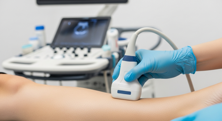
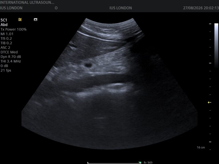
Abdominal aortic aneurysm is primarily diagnosed and monitored using abdominal ultrasound. This non-invasive, radiation-free imaging modality is highly effective, readily available, and cost-efficient. During an ultrasound scan, a transducer emits high-frequency sound waves that penetrate the abdominal wall and reflect off internal structures, including the aorta. These reflected sound waves are then converted into real-time images on a monitor, allowing for precise measurement of the aortic diameter and identification of any aneurysmal dilation. The procedure is painless and typically takes only 10-15 minutes.

Detailed Walk-through of the Patient Experience During an AAA Scan

#### Before the Scan: Preparation

Patients are usually advised to fast for at least 6-8 hours before an abdominal ultrasound. This is because food and liquids can introduce gas into the intestines, which can obscure the view of the aorta and make accurate measurements difficult. Patients should continue to take their regular medications with a small amount of water. It's also helpful to wear loose, comfortable clothing. Upon arrival at the clinic or hospital, patients will typically check in and may be asked to complete a brief questionnaire regarding their medical history and any symptoms.

#### During the Scan: The Procedure

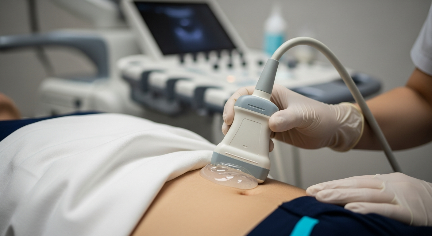
Once called for their scan, the patient will be asked to lie on their back on an examination couch. The sonographer (a specially trained healthcare professional who performs ultrasound scans) will explain the procedure. A clear, water-based gel will be applied to the patient's abdomen. This gel helps the transducer make good contact with the skin and allows the sound waves to travel effectively. The sonographer will then gently press the transducer against the abdomen, moving it slowly across different areas to visualise the entire length of the abdominal aorta. Patients may feel a slight pressure from the transducer, but it should not be painful. They might be asked to hold their breath briefly at certain points to improve image clarity. The sonographer will take several measurements of the aorta's diameter, particularly at its widest point if an aneurysm is present.

#### After the Scan: Results and Next Steps

After the scan is complete, the gel will be wiped off, and the patient can get dressed. The sonographer will not usually provide immediate results, as the images need to be reviewed and interpreted by a radiologist or vascular specialist. The results are typically sent to the referring doctor within a few days. If an AAA is detected, the report will specify its size and any recommendations for follow-up. The referring doctor will then discuss these results with the patient, explain their significance, and outline the recommended surveillance schedule or treatment plan.

Surveillance Schedule Based on Aneurysm Size

The frequency of surveillance scans is meticulously determined by the aneurysm's maximum diameter, reflecting a balance between monitoring growth and avoiding unnecessary procedures. Adherence to established guidelines, such as those from the NHS AAA Screening Programme and international vascular societies, is critical for optimal patient management:

  • Small Aneurysms (3.0–4.4 cm): For aneurysms in this size range, the risk of rupture is relatively low, but continuous monitoring is essential. Annual surveillance scans are recommended. This allows for the detection of any significant growth over a 12-month period, ensuring that intervention can be planned if the aneurysm progresses towards a larger, higher-risk category.
  • Medium Aneurysms (4.5–5.4 cm): As the aneurysm approaches 5.5 cm, the risk of rupture begins to increase more substantially. Therefore, scans every three months are advised for this group. This more frequent monitoring allows for closer observation of the growth rate, enabling prompt decision-making regarding potential intervention. Rapid growth (e.g., >0.5 cm in 6 months or >1 cm in a year) within this range may also trigger earlier consideration for specialist referral.
  • Large Aneurysms (≥5.5 cm): An aneurysm reaching or exceeding 5.5 cm in diameter is generally considered to be at a significantly elevated risk of rupture. At this critical threshold, immediate referral to a vascular specialist is mandated. The specialist will conduct a comprehensive assessment, which may include further imaging (e.g., CT angiography) and a discussion of definitive treatment options, such as surgical repair.

These guidelines are based on extensive research and clinical experience, aiming to identify and manage AAAs effectively while minimising patient anxiety and healthcare costs.

What Happens if an AAA Grows Large?

When an AAA reaches a diameter of 5.5 cm or larger, or demonstrates a rapid growth rate (e.g., more than 0.5 cm in six months), the risk of rupture escalates significantly. At this juncture, the benefits of intervention generally outweigh the risks of watchful waiting. The primary goal of treatment is to prevent rupture, which is associated with an extremely high mortality rate. Two main surgical approaches are considered:

1. Open Surgical Repair

This traditional method involves a major abdominal incision to directly access the aorta. The diseased segment of the aorta is clamped, opened, and replaced with a synthetic graft (typically made of Dacron or PTFE), which is then sewn into place. Open repair is a highly effective and durable solution, offering a long-term cure for AAA. However, it is a major operation associated with a longer recovery period, significant post-operative pain, and a higher risk of complications compared to endovascular repair, particularly in patients with multiple comorbidities.

2. Endovascular Aneurysm Repair (EVAR)

EVAR is a less invasive procedure performed through small incisions in the groin. A catheter is guided through the femoral arteries to the site of the aneurysm. A stent-graft, a fabric tube supported by a metal mesh, is then deployed within the aneurysm to reinforce the weakened aortic wall and exclude the aneurysm from the blood flow. Blood then flows through the graft, bypassing the aneurysm sac, which eventually shrinks. EVAR offers several advantages, including smaller incisions, less pain, shorter hospital stays, and faster recovery. However, it requires lifelong surveillance with imaging scans (CT scans) to monitor for potential complications such as endoleaks (blood leaking back into the aneurysm sac) or graft migration, which may necessitate further interventions.

The choice between open repair and EVAR depends on various factors, including the patient's overall health, the anatomy of the aneurysm, and the expertise of the surgical team. A thorough discussion with a vascular specialist is crucial to determine the most appropriate treatment strategy.

From Our Practice: Anonymised Clinical Scenarios

Understanding AAA surveillance through real-world examples can provide valuable insights into the clinical decision-making process and patient journeys. Here are a few anonymised scenarios from our practice:

Scenario 1: The Vigilant Retiree

Mr. John (72), a former smoker with a history of well-controlled hypertension, was identified with a 3.8 cm AAA during a routine screening ultrasound. Given his risk factors and the aneurysm size, he was placed on an annual surveillance programme. For three consecutive years, his aneurysm remained stable, measuring between 3.8 cm and 4.0 cm. However, at his fourth annual scan, the aneurysm had grown to 4.6 cm. This significant growth prompted a change in his surveillance schedule to quarterly scans. Over the next six months, the aneurysm showed further progression, reaching 5.1 cm. Following a detailed discussion with our vascular specialist, Mr. John opted for an EVAR procedure, which was successfully performed. He recovered well and continues to attend his post-EVAR surveillance appointments, grateful for the early detection and proactive management that prevented a potentially life-threatening event.

Scenario 2: The Unexpected Finding

Mrs. Sarah (68) presented to her GP with non-specific abdominal discomfort. During an unrelated abdominal ultrasound to investigate her symptoms, a 5.6 cm AAA was incidentally discovered. Mrs. Sarah had no known risk factors for AAA, other than her age, and was understandably surprised by the diagnosis. Due to the critical size of the aneurysm, she was immediately referred to our vascular surgery team. After comprehensive imaging and assessment, she underwent open surgical repair. Her recovery was challenging but ultimately successful. This case highlights the often asymptomatic nature of AAAs and the importance of incidental findings in preventing rupture.

Scenario 3: The Rapid Progressor

Mr. David (65), a heavy smoker, was diagnosed with a 4.2 cm AAA. Despite counselling on smoking cessation, he struggled to quit. His initial annual surveillance showed a stable aneurysm. However, at his second annual scan, the aneurysm had rapidly expanded to 4.9 cm. This accelerated growth rate, coupled with his ongoing smoking, raised significant concerns. He was immediately moved to a three-monthly surveillance schedule. Within another three months, the aneurysm measured 5.3 cm. Recognising the imminent danger, Mr. David was strongly advised to undergo intervention. He successfully underwent EVAR and, motivated by the experience, finally committed to smoking cessation, understanding the profound impact it had on his vascular health.

⚠ Red Flags: When to Seek Immediate Medical Attention

While AAAs often remain asymptomatic until they reach a critical size or rupture, it is vital for patients and their families to be aware of potential warning signs. These symptoms can indicate an impending rupture or a rupture that has already occurred, both of which constitute a medical emergency. Seek immediate medical attention by calling emergency services (e.g., 999 in the UK) if you experience any of the following:

* Dizziness or lightheadedness

* Fainting or loss of consciousness

* Pale, clammy, or sweaty skin

* Rapid and weak pulse

* Rapid breathing

* Nausea and vomiting

  • Sudden, Severe Pain: This is the most common symptom of a ruptured AAA. The pain is typically described as tearing or ripping and can be felt in the abdomen, back, or flank. It may radiate to the groin or legs.
  • Pulsating Sensation: A persistent, deep, and often painful pulsating sensation in the abdomen, particularly near the navel, can be a sign of a rapidly expanding aneurysm.
  • Tenderness or Hardness: The abdomen may become tender to touch, or a hard, pulsating mass might be felt, though this is less common and should not be actively sought by palpation by non-medical personnel.
  • Signs of Shock: A ruptured AAA leads to significant internal bleeding, causing hypovolemic shock. Symptoms include:

These symptoms require urgent medical evaluation. Delay in seeking care for a ruptured AAA can have fatal consequences.

Comprehensive FAQs: Common Patient Concerns About AAA Surveillance

Q1: Is an AAA always dangerous?

Not necessarily. Small AAAs (less than 4.5 cm) are generally considered low risk for rupture and are primarily monitored. The danger increases significantly as the aneurysm grows larger, particularly once it reaches 5.5 cm or more. Regular surveillance helps to identify when an aneurysm becomes dangerous and requires intervention.

Q2: What can I do to prevent an AAA from growing?

While you cannot reverse an existing aneurysm, you can significantly slow its growth and reduce your overall cardiovascular risk. Key lifestyle modifications include:

  • Smoking Cessation: This is the single most important step. Quitting smoking dramatically reduces the rate of aneurysm growth.
  • Blood Pressure Control: Meticulous management of high blood pressure through medication and lifestyle changes is crucial.
  • Cholesterol Management: Lowering high cholesterol levels through diet and statin medications can help prevent atherosclerosis progression.
  • Healthy Diet and Exercise: Maintaining a healthy weight and engaging in regular physical activity supports overall cardiovascular health.
  • Diabetes Management: If you have diabetes, keeping blood sugar levels well-controlled is important.

Q3: Are there any alternatives to ultrasound for AAA surveillance?

While ultrasound is the primary and preferred method due to its safety, cost-effectiveness, and non-invasiveness, other imaging modalities may be used in specific circumstances. Computed Tomography (CT) scans, particularly CT angiography, provide more detailed anatomical information and are often used for pre-operative planning or when ultrasound images are inconclusive. However, CT scans involve radiation exposure and are typically reserved for situations where their benefits outweigh these considerations.

Q4: What are the risks of AAA surgery (Open Repair vs. EVAR)?

Both open surgical repair and EVAR are major procedures with potential risks. Open repair carries risks associated with major surgery, including bleeding, infection, heart attack, stroke, kidney problems, and a longer recovery time. EVAR is less invasive but has its own set of risks, such as endoleaks (blood leaking around the stent-graft), graft infection, graft migration, and the need for re-intervention. Your vascular specialist will discuss these risks in detail, tailoring the explanation to your individual health profile.

Q5: How accurate are ultrasound measurements for AAA?

Abdominal ultrasound is highly accurate for measuring the maximum diameter of an AAA, especially when performed by experienced sonographers. However, factors such as bowel gas, patient body habitus, and operator technique can sometimes influence image quality. In cases of uncertainty or when precise anatomical detail is required (e.g., for surgical planning), a CT scan may be performed to confirm measurements and provide additional information.

Q6: Will I need lifelong surveillance after AAA repair?

Yes, lifelong surveillance is typically recommended after both open surgical repair and EVAR. For open repair, follow-up is usually less frequent, focusing on ensuring the integrity of the repair and monitoring for any new aneurysms. For EVAR, surveillance is more rigorous, often involving annual CT scans, to detect and manage potential complications like endoleaks or graft issues that could lead to future rupture. Adherence to this follow-up schedule is crucial for long-term success.

Call to Action (CTA)

If you have concerns about your risk of developing an Abdominal Aortic Aneurysm, have a family history of the condition, or have been diagnosed with an AAA and require expert management, we encourage you to consult with your General Practitioner or a dedicated vascular specialist. Early detection and a meticulously planned surveillance strategy are fundamental to preventing life-threatening complications. Our team at IUS London is committed to providing comprehensive, patient-centred care, utilising the latest diagnostic techniques and evidence-based treatment protocols to ensure your vascular health is optimally managed. Contact us today to discuss your personalised surveillance and management plan.

References

  • NHS Abdominal Aortic Aortic Aneurysm Screening Programme: [Link to NHS website or relevant guideline] – Provides comprehensive information on the UK's national screening programme, including eligibility criteria and surveillance protocols.
  • European Society for Vascular Surgery (ESVS) Clinical Practice Guidelines: [Link to ESVS guidelines] – Offers detailed, evidence-based recommendations for the diagnosis and management of abdominal aortic aneurysms.
  • American Heart Association (AHA) / American College of Cardiology (ACC) Guidelines: [Link to AHA/ACC guidelines] – Provides clinical guidelines for the management of patients with abdominal aortic aneurysms, covering risk assessment, surveillance, and intervention.
  • Journal of Vascular Surgery: [Link to a relevant journal article or journal homepage] – A leading peer-reviewed journal publishing cutting-edge research and clinical reviews on vascular diseases, including AAA.
  • The Lancet: [Link to a relevant article or journal homepage] – A highly respected general medical journal that frequently publishes significant research on cardiovascular health and disease epidemiology.

Book Your Private Ultrasound Scan in London

Same-day appointments available in Kensington. No GP referral required. HCPC-registered consultant sonographers.