Sudden Calf Swelling or Pain — DVT or Strain?

Sudden Calf Swelling or Pain: Is it DVT or a Muscle Strain?

Experiencing sudden swelling or pain in your calf can be alarming and often prompts immediate concern. While frequently attributed to a benign muscle strain, these symptoms can sometimes mask a more serious and potentially life-threatening condition known as Deep Vein Thrombosis (DVT). Understanding the nuanced differences between these conditions, recognising their distinct symptoms, and knowing precisely when to seek urgent medical attention are not merely advisable but absolutely crucial for safeguarding your health and preventing severe complications.

⚠ Red Flags: When to Seek Immediate Medical Attention

It is imperative to recognise that certain symptoms warrant immediate and decisive action. If you experience any of the following, do not delay; seek immediate Accident & Emergency (A&E) attention without hesitation:

  • Sudden, severe calf pain without obvious injury: Unlike muscle strains that typically follow a specific incident or exertion, DVT pain can emerge without a clear precipitating event, often described as a persistent ache or cramping sensation.
  • Significant swelling in one calf: DVT usually affects one leg, leading to noticeable swelling that is often greater than any swelling in the other leg. This unilateral swelling is a key differentiator from more generalised conditions.
  • Warmth or redness over the affected area: The presence of a blood clot can cause inflammation, leading to the skin over the affected vein feeling warm to the touch and appearing reddish or discoloured.
  • Shortness of breath, chest pain, or lightheadedness (possible pulmonary embolism): These are critical warning signs of a pulmonary embolism (PE), a life-threatening complication where a part of the DVT clot breaks off and travels to the lungs. A PE requires emergency medical intervention.

Understanding the Causes of Calf Swelling and Pain

Calf pain and swelling can stem from a variety of causes, ranging from the relatively benign to the critically serious. Accurate diagnosis is paramount for appropriate treatment.

Deep Vein Thrombosis (DVT)

Deep Vein Thrombosis (DVT) occurs when a blood clot (thrombus) forms in one of the deep veins, most commonly in the legs, but it can also occur in the arms or other parts of the body. These clots can partially or completely block blood flow, leading to pain, swelling, and warmth. The primary danger of DVT lies in the potential for a piece of the clot to break off, travel through the bloodstream, and lodge in the lungs, causing a pulmonary embolism (PE). This is a medical emergency that can be fatal. Ultrasound is the primary diagnostic tool for DVT, offering a non-invasive and highly accurate method for visualising the veins and detecting clots.

Baker's Cyst (Popliteal Cyst)

A Baker's Cyst, also known as a popliteal cyst, is a fluid-filled sac that forms behind the knee. It is typically caused by an accumulation of joint fluid (synovial fluid) that has been pushed out of the knee joint due to underlying conditions such as arthritis or cartilage tears. While often asymptomatic, a Baker's cyst can cause pain, stiffness, and swelling behind the knee. If the cyst ruptures, the fluid can leak into the calf, mimicking DVT symptoms with sudden pain and swelling. Ultrasound is highly effective in diagnosing Baker's cysts, differentiating them from other causes of calf discomfort, and assessing for rupture.

Muscle Strain

A muscle strain, commonly referred to as a pulled muscle, occurs when muscle fibres are stretched or torn due to overexertion, sudden movements, or trauma. This is a very common injury, particularly among athletes or individuals engaging in physical activity. Symptoms typically include sudden, sharp pain during or immediately after the activity, tenderness, bruising, and localised swelling. While generally less severe than DVT, a significant muscle tear can still cause considerable discomfort and limit mobility. Ultrasound can confirm the diagnosis of a muscle strain, assess the extent of the injury, and rule out other conditions.

Risk Factors for DVT — Who Is Most at Risk?

Deep Vein Thrombosis is not a random occurrence; certain factors significantly increase an individual's susceptibility to developing blood clots. Understanding these risk factors is crucial for prevention and early detection. The primary risk factors for DVT can be broadly categorised as conditions that promote blood stasis (slow blood flow), hypercoagulability (increased tendency for blood to clot), or endothelial injury (damage to the blood vessel lining).

  • Immobility: Prolonged periods of inactivity, such as extended bed rest after surgery, hospitalisation, or long journeys (e.g., flights, car rides), significantly slow blood flow in the veins. When muscles are not contracting, they cannot effectively pump blood back to the heart, leading to blood pooling in the lower extremities and increasing the risk of clot formation.
  • Recent Surgery: Surgical procedures, particularly major operations involving the abdomen, pelvis, hip, or knee, are significant risk factors. Surgery can cause direct damage to blood vessels, trigger an inflammatory response that promotes clotting, and often necessitates periods of immobility during recovery. Anaesthesia can also contribute to reduced blood flow.
  • Long-Haul Travel: Extended periods of sitting in confined spaces, common during long-distance flights or car journeys lasting more than four hours, can lead to blood stasis in the legs. The cramped conditions and reduced movement hinder the calf muscle pump, increasing the risk of DVT, often referred to as economy class syndrome.
  • Pregnancy and Postpartum Period: Pregnancy increases the risk of DVT due to several physiological changes. The growing uterus puts pressure on the pelvic veins, impeding blood flow from the legs. Additionally, hormonal changes (increased oestrogen) make the blood more prone to clotting, a natural protective mechanism to prevent excessive bleeding during childbirth. This elevated risk persists for up to six weeks postpartum.
  • Oral Contraceptives and Hormone Replacement Therapy (HRT): Medications containing oestrogen, such as oral contraceptive pills and hormone replacement therapy, can increase the blood's clotting ability. Oestrogen can affect the production of clotting factors, making the blood more hypercoagulable and thus elevating the risk of DVT.
  • Thrombophilia (Inherited or Acquired Clotting Disorders): Thrombophilia refers to a group of conditions where the blood has an increased tendency to clot. These can be inherited (e.g., Factor V Leiden mutation, Prothrombin gene mutation) or acquired (e.g., Antiphospholipid Syndrome). Individuals with thrombophilia have a significantly higher baseline risk of DVT, especially when combined with other risk factors.
  • Obesity: Being overweight or obese is a well-established risk factor for DVT. Excess weight places increased pressure on the veins in the pelvis and legs, hindering blood flow. Obesity is also associated with chronic inflammation and changes in clotting factors, further contributing to a pro-thrombotic state.

What Happens During a DVT Ultrasound Scan at IUS London

At IUS London, a Deep Vein Thrombosis (DVT) ultrasound scan is a crucial, non-invasive diagnostic procedure performed by highly skilled sonographers to accurately assess your veins and detect the presence of blood clots. Our state-of-the-art equipment ensures precise imaging and a thorough evaluation.

The Compression Ultrasound Technique

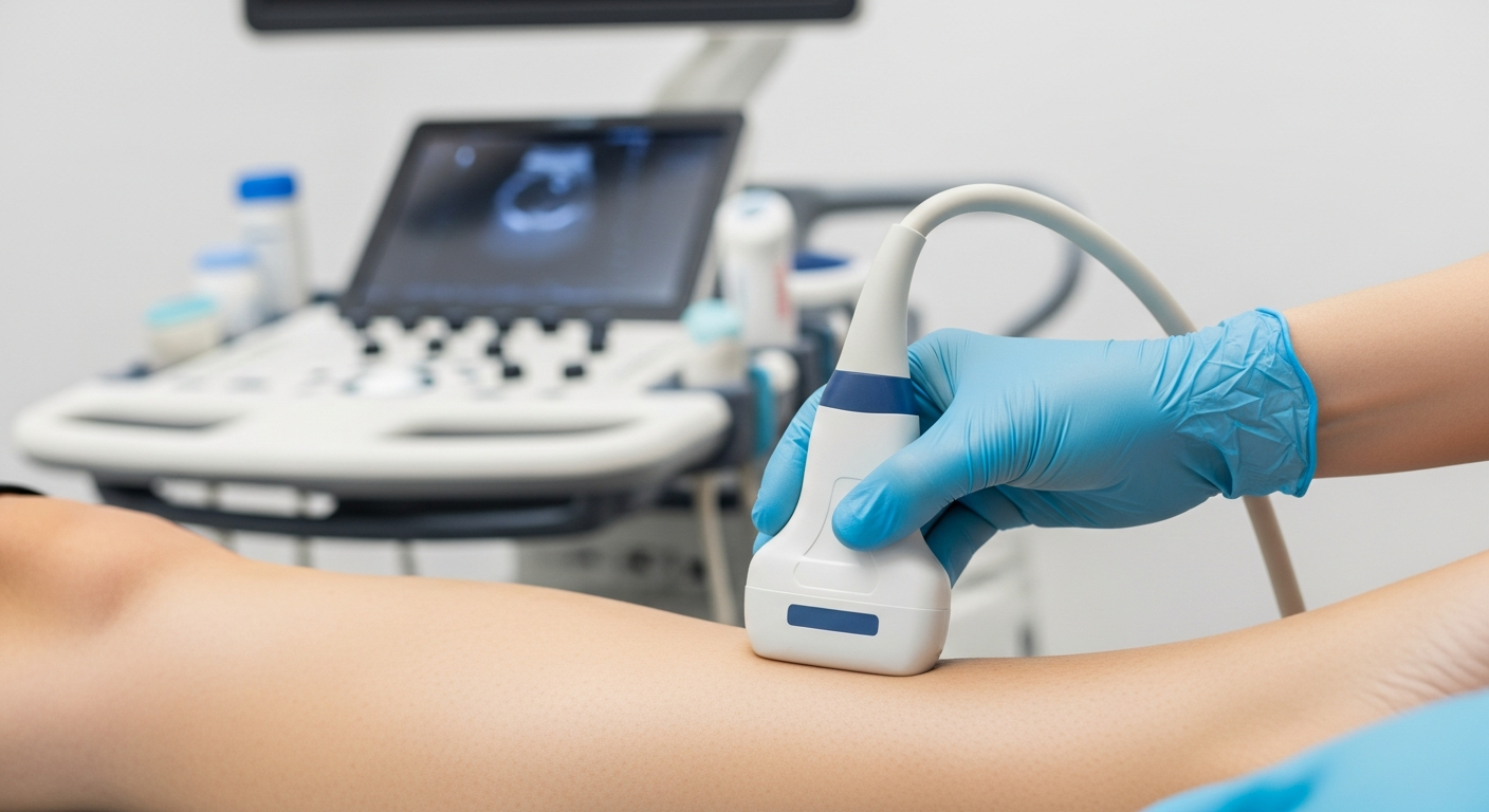
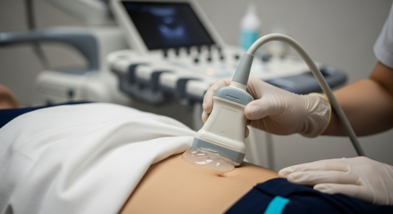
The cornerstone of DVT diagnosis via ultrasound is the compression ultrasound technique. During this procedure, you will typically lie comfortably on an examination couch. The sonographer will apply a small amount of warm gel to your skin over the areas to be examined, usually starting from your groin and extending down to your calf. A handheld transducer (probe) is then gently pressed against your skin. The key principle here is that healthy veins are easily compressible; they flatten when pressure is applied. If a blood clot is present within a vein, it will prevent the vein from compressing fully, indicating the presence of a thrombus. The sonographer systematically examines various segments of the deep venous system, including the common femoral vein, superficial femoral vein, popliteal vein, and calf veins, applying gentle compression at regular intervals.

Doppler Assessment

In addition to the compression technique, Doppler assessment is an integral part of a comprehensive DVT ultrasound. Doppler ultrasound uses sound waves to evaluate blood flow within the veins. This technique allows the sonographer to:

  • Visualise blood flow: Observe the direction and velocity of blood flow.
  • Detect obstructions: Identify areas where blood flow is absent or significantly reduced, which can indicate a clot.
  • Assess venous patency: Confirm that the veins are open and blood is flowing freely.

By combining real-time imaging with Doppler flow analysis, our sonographers can gain a complete picture of your venous health, identifying any abnormalities that may suggest DVT.

What the Report Includes

Following your DVT ultrasound scan at IUS London, a detailed report will be compiled by our specialist sonographer and reviewed by a consultant radiologist. This comprehensive report will include:

  • Clinical Findings: A summary of the sonographer's observations during the scan.
  • Presence or Absence of DVT: A clear statement regarding whether a DVT was identified.
  • Location and Extent of Clot (if present): Precise details on where any clots are located and how much of the vein they affect.
  • Vein Compressibility: Documentation of the compressibility of the deep veins.
  • Doppler Flow Characteristics: Description of blood flow patterns and any abnormalities detected.
  • Other Findings: Any other relevant observations, such as the presence of a Baker's cyst or muscle injury, which might explain your symptoms.
  • Recommendations: Where appropriate, recommendations for further management or follow-up will be provided. This report is then promptly shared with you and your referring clinician to ensure timely and informed decision-making regarding your care.

Comparative Analysis of Calf Conditions

FeatureDVTBaker's Cyst (Ruptured)Muscle Strain
OnsetSudden, without injury, or gradualSudden if ruptured, often preceded by knee painDuring or immediately after physical activity
PainAching, cramping, tenderness, often worse with standing or walkingSharp, sudden pain if ruptured, can be diffuse in the calfSharp, tearing, localised pain, worse with movement or palpation
SwellingUnilateral, significant, often pitting oedemaDiffuse in the calf, can be significantLocalised to the injured muscle, sometimes with bruising
Skin ChangesWarmth, redness, discolouration (bluish or reddish-purple)May have some redness or warmth due to inflammationMay have bruising, tenderness to touch
Associated SymptomsTenderness along the vein, fever (rare), dilated superficial veinsKnee stiffness, limited range of motion in the kneeMuscle weakness, difficulty bearing weight on the affected leg

From Our Practice

At IUS London, we frequently encounter patients presenting with acute calf pain and swelling who initially dismissed symptoms as a minor strain. Our clinical experience highlights the critical importance of accurate and timely diagnosis. We have seen numerous cases where early intervention following a DVT diagnosis, confirmed by our advanced ultrasound scans, has prevented serious complications such as pulmonary embolism. Conversely, we also provide reassurance to many patients whose symptoms are ultimately attributed to less severe conditions like muscle strains or ruptured Baker's cysts, allowing them to pursue appropriate, targeted treatment and recovery plans. Our commitment is to provide clarity and peace of mind through expert diagnostic imaging.

Book Your Private Ultrasound Scan

If you are experiencing sudden calf swelling or pain, or have concerns about DVT, do not hesitate to book a private ultrasound scan at IUS London. Our experienced team is dedicated to providing rapid, accurate diagnosis and comprehensive care. Early detection is key to effective management and preventing potential complications. Book Now

References

[1] Bhatt, M., et al. (2020). Diagnosis of deep vein thrombosis. Blood advances, 4(17), 4190-4200.

[2] Kraaijpoel, N., et al. (2020). Diagnostic accuracy of three ultrasonography strategies for DVT. PLoS ONE, 15(1), e0227310.

[3] Bright, J., et al. (2017). Ultrasound Diagnosis of Calf Injuries. Sports Health, 9(4), 352-357.

[4] Pagan-Rosado, R., et al. (2025). Calf Strains in Athletes. Sports Medicine - Open, 11(1), 1-12.

[5] Anderson Jr, F. A., et al. (2003). Risk Factors for Venous Thromboembolism. Circulation, 107(23 Suppl 1), I-9-I-15.

[6] Waheed, S. M., et al. (2023). Deep Venous Thrombosis. StatPearls Publishing.

[7] Needleman, L., et al. (2018). Ultrasound for Lower Extremity Deep Venous Thrombosis. Circulation, 137(14), 1505-1515.

[8] Ibrahim, M. Z., et al. (2020). Doppler Ultrasonographic Evaluation of Lower Limbs Deep-Vein Thrombosis. Journal of Clinical Imaging Science, 10, 15.

Book Your Private Ultrasound Scan in London

Same-day appointments available in Kensington. No GP referral required. HCPC-registered consultant sonographers.