Vascular Disease — Conditions of the Arteries and Veins
What goes wrong in blood vessels, which conditions ultrasound can assess, and which need a different test entirely.

Vascular disease covers anything affecting the blood vessels outside the heart, and it divides cleanly into two groups that behave quite differently. Arterial disease is a supply problem — vessels narrow, harden or dilate, and tissue downstream is starved. Venous disease is a drainage problem — blood struggles to get back, pools, or clots.
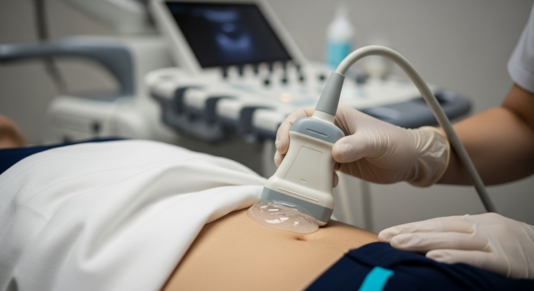
Ultrasound with Doppler is the first-line test for much of this, because it shows both the vessel wall and the blood moving through it in real time, without radiation or contrast. It is not the right test for everything, and this page is explicit about where it stops.
A limb that is suddenly painful, pale, cold, numb or weak — acute loss of arterial supply is measured in hours. Sudden severe abdominal or back pain with faintness, which can indicate a ruptured aneurysm. Sudden breathlessness or chest pain alongside a swollen calf, suggesting clot that has travelled to the lungs. Also seek same-day assessment for a newly swollen, hot, painful calf — possible DVT needs scanning quickly, and NHS pathways exist specifically for it.
Conditions affecting the arteries
Peripheral arterial disease. Narrowing from atherosclerosis, typically in the legs. The classic symptom is cramping calf pain on walking that eases with rest. Doppler measures flow velocities and waveform shape to locate and grade narrowings.
Carotid artery stenosis. Narrowing of the neck arteries supplying the brain, an important stroke risk factor and often symptomless until it is not. Ultrasound grades it by velocity measurement, which is why the carotid scan is a measurements examination rather than a picture-taking one.
Abdominal aortic aneurysm. Dilation of the main abdominal artery, usually symptomless until rupture, and readily measured on ultrasound — which is why AAA scanning is one of the clearest cases for imaging someone without symptoms.
Buerger’s disease (thromboangiitis obliterans). Inflammation and clotting in the small and medium arteries of the hands and feet, strongly associated with tobacco use and typically affecting younger smokers. Ultrasound shows reduced or absent flow in the distal vessels and helps exclude atherosclerotic disease and embolism as alternative explanations. Diagnosis is clinical, supported by imaging — and stopping smoking completely is the single intervention that changes its course.
Takayasu arteritis. A rare large-vessel vasculitis affecting the aorta and its main branches, most often in younger women, causing wall thickening and narrowing. Ultrasound can demonstrate the characteristic uniform wall thickening in accessible vessels such as the carotids and subclavians, though full assessment uses CT or MR angiography and inflammatory blood markers. It is treatable, generally with immunosuppression, and managed by a rheumatology or vascular specialist.
Conditions affecting the veins
Deep vein thrombosis. A clot in a deep vein, usually of the leg, presenting with swelling, warmth and pain. Ultrasound is the definitive test: a normal vein compresses under gentle probe pressure, and a vein that will not compress contains clot. That simple manoeuvre is the core of a DVT scan.
Chronic venous insufficiency and varicose veins. Valves that should keep blood moving upward fail, so blood refluxes and pools — producing aching, swelling, skin changes around the ankle and, over time, ulceration. Doppler identifies exactly which valves are incompetent and where, which is what any treatment planning depends on.
Superficial thrombophlebitis. Clot with inflammation in a surface vein, felt as a tender cord under the skin. Usually less serious than DVT, but scanning matters because it can extend into the deep system.
Post-thrombotic syndrome. Persistent swelling, heaviness and skin changes following a previous DVT, caused by lasting valve damage.
Matching the symptom to the examination
| What you have noticed | Usual examination |
|---|---|
| Swollen, hot, painful calf — suspected clot | DVT scan — £179 (seek same-day assessment first) |
| Aching, heavy legs, visible varicose veins, ankle skin changes | Venous insufficiency assessment |
| Stroke risk review, or a neck bruit heard by your doctor | Carotid scan — £189 |
| Aneurysm screening, or a family history of one | AAA scan — £139 |
| Cramping calf pain on walking that eases with rest | See your GP — arterial assessment usually starts with ankle-brachial pressure measurement |
What ultrasound cannot do here
Vessels it cannot reach. Sound does not travel through bone or gas, so the intracranial arteries, the coronary arteries and parts of the chest are not assessable by ordinary ultrasound. Bowel gas can also obscure the abdominal vessels on a given day.
Whole-map assessment. Ultrasound examines vessels one segment at a time. Where a complete map is needed — before surgery, or in extensive disease — CT or MR angiography does what ultrasound cannot.
Inflammatory activity. In vasculitis, ultrasound shows wall changes but not the level of inflammation. Blood markers and sometimes PET imaging are used for that.
Our guide to ultrasound versus MRI and CT sets out where each modality belongs, and how Doppler works explains the colours and waveforms these examinations rely on.
Your questions answered
What is a vascular ultrasound scan?
How does a scan detect a DVT?
Can ultrasound diagnose Buerger’s disease?
Can ultrasound detect Takayasu arteritis?
What is the difference between arterial and venous disease?
Do I need a referral for a vascular scan?
When is ultrasound not the right test?
What we offer
Vascular scanning in Kensington
Arterial and venous Doppler by HCPC-registered sonographers, measurements explained on screen, and a written report usually within two hours for your GP or vascular specialist. No referral needed.
Kensington, London W8 4ED
3 mins from Notting Hill Gate
This article is general information, not individual medical advice. Suspected deep vein thrombosis, sudden limb ischaemia, suspected aneurysm rupture or symptoms of pulmonary embolism are emergencies requiring immediate NHS assessment. Vasculitis and arterial disease are managed by specialist teams; imaging supports diagnosis but does not replace clinical assessment.
Written and clinically reviewed by the HCPC-registered sonographers who perform this scan at IUS London — a CQC-registered diagnostic ultrasound clinic (Provider ID 1-2775844974). Your own findings are explained to you at the scan and set out in your report.
Author: Yianni Kiromitis, Senior Sonographer, HCPC RA38415
Medically reviewed: 4 August 2026