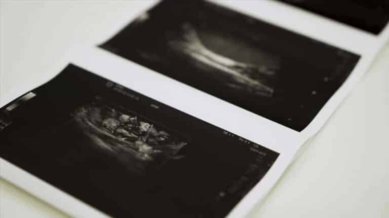
Gynaecological Ultrasound — A Patient’s Guide
What a gynae scan is, how it is performed, every condition it can identify, and how to read what the report says afterwards.

A gynaecological ultrasound — also called a gynae scan, gynaecology scan or pelvic ultrasound — is an examination of the female reproductive organs using sound waves. It looks at the uterus (womb), the endometrium (the lining of the womb), both ovaries, and the surrounding areas of the pelvis. It uses no radiation, causes no lasting effects, and produces live images that can be explained to you as they appear.
It is the first-line investigation for most gynaecological symptoms — pelvic pain, heavy or irregular bleeding, suspected cysts or fibroids, bleeding after menopause, and fertility assessment — because it answers a large proportion of those questions in a single 30-minute appointment.
How the scan is performed
There are two approaches, and most gynaecological scans use one or both.
Transabdominal — the probe passes over the lower abdomen with gel. It requires a comfortably full bladder, which lifts the bowel out of the way and creates a window through to the uterus. It gives a wide overview and is the better approach for a very large uterus or a big ovarian cyst that extends beyond the pelvis.
Transvaginal — a slim probe is placed in the vagina, giving a much closer and more detailed view. It needs an empty bladder, and it is the more informative approach for the endometrium, the ovaries and early pregnancy. It is entirely your choice, and our guide to transvaginal scanning covers consent, comfort and what to expect — with a first-time guide if you have not had one before.
Using both is common: the abdominal view establishes the overall picture, the transvaginal view supplies the detail. An honest report states which was used and anything that could not be assessed.
Conditions a gynaecological ultrasound can identify
| Condition | What the scan contributes |
|---|---|
| Fibroids | Confirms them, counts and measures each, and locates them — location determines whether they explain your symptoms |
| Ovarian cysts | Distinguishes simple functional cysts from those needing follow-up, using size, contents and blood flow |
| Polycystic ovarian appearance | Identifies the characteristic pattern of many small follicles — an appearance, which becomes PCOS only with clinical criteria |
| Endometrial thickening or polyps | Measures the lining against your cycle stage or menopausal status |
| Adenomyosis | Suggests it from the appearance of the uterine wall, though MRI is sometimes needed for certainty |
| Pelvic infection complications | Detects abscess or fluid-filled tubes — though mild infection often looks normal |
| Endometriosis | Identifies endometriomas on the ovaries; superficial disease is frequently not visible, so a normal scan does not exclude it |
| Coil (IUD) position | Confirms the device sits correctly within the cavity |
| Early pregnancy | Confirms location and viability — including excluding an ectopic pregnancy |
Two honest limits worth knowing. Ultrasound cannot diagnose infection directly, and it cannot exclude endometriosis — superficial disease is often invisible on any scan. And no ultrasound diagnoses cancer; it identifies features that require tissue diagnosis to resolve.
Why a gynae scan gets arranged
- Pelvic pain — persistent, cyclical, or sudden and severe
- Heavy, prolonged or irregular periods
- Bleeding after the menopause — which always warrants assessment
- Bleeding between periods or after sex
- Difficulty conceiving, or monitoring during fertility treatment
- A suspected cyst or fibroid found on examination
- Checking a coil where threads cannot be felt
- Following up a previous finding to see whether it has changed
Preparation, timing and the appointment
Preparation. For a transabdominal scan, drink around a litre of water an hour beforehand and do not empty your bladder. For a transvaginal scan, an empty bladder is more comfortable. Where both may be used, arrive with a full bladder — you can empty it before the internal part. No fasting is required.
Timing in your cycle. Often it does not matter, but sometimes it does. Scanning in the first half of the cycle, soon after a period, gives the clearest view of the womb lining and is preferred when the lining or possible polyps are the question. Fertility scans are timed deliberately to the cycle. You can be scanned during a period — it is safe and routine.
The appointment takes about 30 minutes. Findings are explained on screen as they are found, and a written report follows — usually within two hours — written so your GP or gynaecologist can act on it. Our guide to reading a pelvic report decodes the terminology, from endometrial thickness to free fluid in the pouch of Douglas.
Your questions answered
What is a gynaecological ultrasound?
What is the difference between a gynae scan and a pelvic ultrasound?
Does a gynaecologist perform the scan?
What can a gynaecological ultrasound detect?
Do I need a full bladder?
Can I have a gynae scan during my period?
Do I need a GP referral?
The conditions in detail
A gynaecological scan, explained as it happens
Transabdominal and transvaginal scanning by HCPC-registered sonographers, findings shown to you on screen, and a written report usually within two hours — no GP referral needed.
Kensington, London W8 4ED
3 mins from Notting Hill Gate
This article is general information, not individual medical advice. Ultrasound findings should be interpreted alongside your symptoms and examination by your GP or gynaecologist. Bleeding after the menopause, or sudden severe pelvic pain, needs prompt medical assessment.
Written and clinically reviewed by the HCPC-registered sonographers who perform this scan at IUS London — a CQC-registered diagnostic ultrasound clinic (Provider ID 1-2775844974). Your own findings are explained to you at the scan and set out in your report.
Author: Yianni Kiromitis, Senior Sonographer, HCPC RA38415
Medically reviewed: 1 August 2026