Introduction
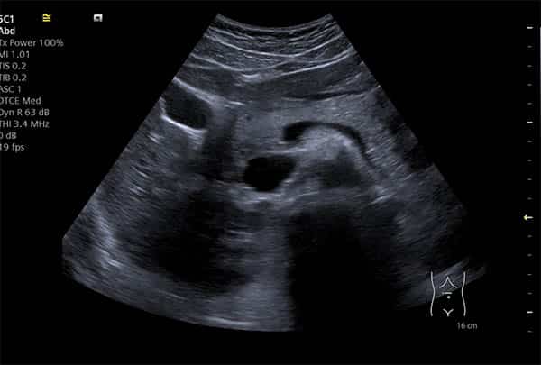
Stomach problems are a common complaint, ranging from mild discomfort to severe, debilitating pain. The abdomen houses a multitude of organs, and pinpointing the exact cause of symptoms can be challenging. Medical imaging plays a crucial role in diagnosing these conditions, and ultrasound is often a first-line investigation due to its non-invasive nature, portability, and lack of radiation exposure. This article explains the capabilities and limitations of abdominal ultrasound in evaluating stomach problems.
⚠ Red Flags (A&E): When to Seek Immediate Medical Attention
While many stomach problems are benign, certain symptoms warrant immediate medical attention at an Accident & Emergency (A&E) department. Recognizing these red flags is crucial for timely intervention and can be life-saving. Seek urgent care if you experience:
- Sudden, severe abdominal pain that is unbearable or rapidly worsening.
- Abdominal pain accompanied by fever, chills, or persistent vomiting.
- Bloody stools, black, tarry stools (melena), or vomiting blood.
- Severe tenderness when touching your abdomen.
- Inability to pass gas or have a bowel movement.
- Jaundice (yellowing of the skin or eyes).
- Sudden, unexplained weight loss.
- Signs of shock, such as rapid heart rate, low blood pressure, confusion, or cold, clammy skin.
These symptoms could indicate serious conditions such as appendicitis, bowel obstruction, perforated ulcer, acute pancreatitis, or ectopic pregnancy, which require prompt diagnosis and treatment.
What is an Abdominal Ultrasound?
An abdominal ultrasound, also known as a sonogram, is a medical imaging technique that uses high-frequency sound waves to create real-time images of organs and structures within the abdomen. A small, handheld device called a transducer is placed on the skin over the abdomen, and it emits sound waves that travel through the body. These sound waves bounce off organs and tissues, and the transducer captures the echoes. A computer then converts these echoes into a visual image on a screen. Unlike X-rays or CT scans, ultrasound does not use ionizing radiation, making it a safe option for repeated examinations, pregnant patients, and children.
What an Abdominal Ultrasound Can See: Diagnostic Capabilities
Abdominal ultrasound is a versatile diagnostic tool capable of visualizing various organs and identifying a range of conditions that cause stomach problems. Its utility is particularly highlighted in emergency settings for acute abdominal pain [1].
Gallbladder and Biliary System
Ultrasound is considered the method of choice for visualizing the gallbladder and identifying gallstones (cholelithiasis) [5]. It can effectively detect:
- Gallstones: Solid particles that form in the gallbladder, often causing pain, especially after fatty meals.
- Cholecystitis: Inflammation of the gallbladder, usually due to gallstones blocking the cystic duct. Ultrasound can show gallbladder wall thickening, pericholecystic fluid, and a positive sonographic Murphy's sign.
- Biliary obstruction: Blockage of the bile ducts, which can be caused by gallstones, tumors, or strictures. Ultrasound can identify dilated bile ducts and sometimes the cause of obstruction.
Liver
The liver is a large organ in the upper right abdomen, and ultrasound can assess its size, shape, and texture. It can detect:
- Fatty liver disease (steatosis): Accumulation of fat in the liver, often associated with obesity, diabetes, and alcohol consumption.
- Liver cysts and masses: Fluid-filled sacs or solid growths, which may be benign or malignant.
- Cirrhosis: Advanced scarring of the liver, often due to chronic liver disease.
- Hepatitis: Inflammation of the liver.
Pancreas
While the pancreas can be challenging to visualize completely due to its retroperitoneal location and overlying bowel gas, ultrasound can still provide valuable information, especially for the head of the pancreas. It can help diagnose:
- Pancreatitis: Inflammation of the pancreas, which can be acute or chronic. Ultrasound may show pancreatic enlargement, fluid collections, or pseudocysts.
- Pancreatic masses: Tumors or other growths.
Kidneys and Urinary Tract
Although primarily associated with urinary symptoms, kidney problems can manifest as abdominal pain. Ultrasound is excellent for evaluating the kidneys and urinary tract:
- Kidney stones (nephrolithiasis): Stones within the kidneys or ureters, causing severe flank or abdominal pain.
- Hydronephrosis: Swelling of the kidney due to urine backup, often caused by obstruction.
- Kidney cysts and masses.
- Bladder abnormalities.
Spleen
Ultrasound can assess the size and condition of the spleen, which can be enlarged (splenomegaly) due to various conditions, including infections, liver disease, and blood disorders.
Appendix
In cases of suspected appendicitis, especially in young females, ultrasound can be a valuable initial imaging modality. It can identify an inflamed, non-compressible appendix, periappendiceal fluid, and appendicoliths. While CT often has higher sensitivity for appendicitis, ultrasound is preferred in certain populations to avoid radiation exposure [2].
Bowel (Gastrointestinal Tract)
While not the primary modality for detailed bowel examination (endoscopy and CT are often preferred), gastrointestinal ultrasound (GIUS) is gaining recognition, particularly in emergency settings for acute abdominal pain [1]. It can be useful for:
- Detecting bowel wall thickening: Indicative of inflammation (e.g., Crohn's disease, ulcerative colitis) or infection.
- Identifying fluid collections or abscesses.
- Diagnosing intussusception: A condition where one segment of the intestine telescopes into another, more common in children.
- Assessing diverticulitis: Inflammation of diverticula in the colon.
Abdominal Aorta
Ultrasound is highly effective in screening for and monitoring abdominal aortic aneurysms (AAA), a potentially life-threatening condition where the aorta weakens and bulges [5].
What an Abdominal Ultrasound Cannot See (Limitations)
Despite its many advantages, abdominal ultrasound has certain limitations that can affect its diagnostic accuracy:
- Bowel Gas: Gas in the intestines can scatter sound waves, making it difficult to visualize structures behind or beneath it. This is a significant challenge for organs like the pancreas and parts of the bowel.
- Obesity: Excess abdominal fat can attenuate sound waves, reducing image quality and penetration, making it harder to obtain clear images in obese patients.
- Operator Dependence: The quality and interpretation of an ultrasound scan are highly dependent on the skill and experience of the sonographer and reporting clinician. This subjectivity can lead to variability in diagnostic accuracy.
- Limited Field of View: Ultrasound provides a relatively narrow field of view compared to CT or MRI, which can make it challenging to assess diffuse or widespread pathologies.
- Inability to See Through Bone: Sound waves cannot penetrate bone, limiting its utility for structures obscured by the ribs or spine.
- Specificity for Certain Conditions: While ultrasound can detect abnormalities, it may not always provide a definitive diagnosis for certain conditions, often requiring further imaging (e.g., CT or MRI) or endoscopic procedures for confirmation [2, 8]. For instance, while it can detect inflammation, distinguishing between different causes of inflammation can be difficult.
- Chronic Abdominal Pain of Unknown Etiology: For chronic upper abdominal pain where conventional imaging (CT, MRI, endoscopy) has yielded no diagnosis, endoscopic ultrasound (EUS) has shown a higher diagnostic yield compared to transabdominal ultrasound, particularly for conditions like CBD stones, gallbladder microlithiasis, and early chronic pancreatitis [3]. This highlights that for persistent, unexplained pain, more specialized ultrasound techniques may be necessary.
Can an Ultrasound Detect Gastritis or H. pylori?
This is one of the questions we are asked most often, and the honest answer matters. Ultrasound is not the test that diagnoses gastritis. Gastritis — inflammation of the stomach lining — is confirmed by gastroscopy (endoscopy), where a camera examines the stomach lining directly and small samples can be taken. On ultrasound, the stomach is a gas-filled organ, and gas blocks sound waves, so the lining cannot be assessed reliably. Occasionally an ultrasound will show thickening of the stomach wall that raises suspicion, but a normal ultrasound does not rule gastritis out, and an abnormal one still needs endoscopy to confirm.
So why scan at all if gastritis is suspected? Because the symptoms of gastritis — upper abdominal pain, discomfort after eating, bloating, nausea — overlap heavily with conditions ultrasound assesses very well: gallstones, biliary problems, fatty liver, pancreatic inflammation and kidney stones. An abdominal ultrasound is often the sensible first step precisely because it can identify or exclude these look-alike causes quickly and without radiation, helping your GP decide whether endoscopy is the right next test.
Can ultrasound detect H. pylori? No. Helicobacter pylori — the bacterium behind many cases of gastritis and stomach ulcers — cannot be seen on any ultrasound scan. It is diagnosed with a urea breath test or a stool antigen test, both usually arranged through your GP, and treated with a course of antibiotics if positive. If your symptoms are persistent, or you have red-flag features such as unintentional weight loss, difficulty swallowing, vomiting blood or black stools, see your GP promptly rather than booking a scan first.
Differential Diagnosis: Comparing Symptoms and Conditions
Abdominal pain is a complex symptom with a vast differential diagnosis. Ultrasound helps narrow down the possibilities by providing visual evidence of organ pathology. Below is a comparison of common symptoms and conditions, illustrating how ultrasound aids in differentiation.
| Symptom/Location of Pain | Possible Conditions | How Ultrasound Helps Differentiate |
|---|---|---|
| **Right Upper Quadrant Pain** | Gallstones, Cholecystitis, Hepatitis, Liver Mass, Pancreatitis | Visualizes gallstones, gallbladder inflammation, liver size/texture, pancreatic inflammation. |
| **Right Lower Quadrant Pain** | Appendicitis, Ovarian Cyst/Torsion, Ectopic Pregnancy, Crohn's Disease, Kidney Stone | Identifies inflamed appendix, ovarian abnormalities, fluid in pelvis, kidney stones/hydronephrosis. |
| **Left Upper Quadrant Pain** | Splenomegaly, Pancreatitis, Gastritis, Kidney Stone | Assesses spleen size, pancreatic inflammation, kidney stones/hydronephrosis. |
| **Left Lower Quadrant Pain** | Diverticulitis, Ovarian Cyst/Torsion, Ectopic Pregnancy, Ulcerative Colitis, Kidney Stone | Detects bowel wall thickening (diverticulitis), ovarian abnormalities, kidney stones/hydronephrosis. |
| **Epigastric Pain** | Gastritis, Peptic Ulcer Disease, Pancreatitis, Gallstones | Can show pancreatic inflammation, gallstones. Less direct for gastritis/ultrasound. |
| **Generalized Abdominal Pain** | Gastroenteritis, Bowel Obstruction, Peritonitis, Mesenteric Ischemia | Can identify dilated bowel loops (obstruction), free fluid (peritonitis), bowel wall changes. |
From Our Practice at IUS London
At IUS London, we frequently encounter patients presenting with non-specific upper abdominal discomfort, often exacerbated by fatty foods. A common clinical pattern involves a patient, typically a female in her late 30s to 50s, reporting intermittent right upper quadrant pain, sometimes radiating to the back, accompanied by bloating and indigestion. Initial general practitioner assessment might suggest dyspepsia. However, our high-resolution abdominal ultrasound often reveals subtle findings such as gallbladder sludge or small gallstones that were not detected by previous imaging or were dismissed as insignificant. In one anonymised case, a 45-year-old woman presented with recurrent post-prandial pain. Her GP had prescribed antacids, but symptoms persisted. Our ultrasound identified multiple small gallstones and mild gallbladder wall thickening, leading to a diagnosis of chronic cholecystitis. This finding allowed for appropriate referral to a surgeon for cholecystectomy, resolving her long-standing symptoms. This highlights the importance of detailed, high-quality ultrasound imaging in uncovering the underlying cause of seemingly vague abdominal complaints.
Why Choose IUS London for Your Ultrasound Scan?
At IUS London, we pride ourselves on providing a premium, patient-centred experience with unparalleled diagnostic accuracy. Our clinic in Kensington is equipped with modern ultrasound technology, operated by highly experienced and compassionate sonographers and consultant radiologists. We understand the anxiety that can accompany stomach problems, and we are committed to offering rapid access to scans, detailed explanations, and prompt results. Our focus on clinical excellence ensures that you receive the most thorough and accurate assessment, guiding you towards the right treatment pathway.
Conclusion
Abdominal ultrasound is an invaluable tool in the diagnostic arsenal for stomach problems, offering a safe, non-invasive, and effective way to visualize many abdominal organs. While it excels in detecting conditions of the gallbladder, liver, kidneys, and can provide crucial insights into acute abdominal pain, it does have limitations, particularly concerning bowel gas and the need for operator expertise. Understanding both its capabilities and limitations is key to its appropriate use in clinical practice. For accurate and timely diagnosis of your stomach concerns, a high-quality ultrasound performed by experienced professionals is paramount.
References
1. Boccatonda, A., et al. (2023). Gastrointestinal Ultrasound in Emergency Setting. Journal of Clinical Medicine.
https://consensus.app/papers/details/6d4fb49fe19b5690bb7513f9aa6f1c4a/?utm_source=unknown
2. Randen, A., et al. (2011). A comparison of the Accuracy of Ultrasound and Computed Tomography in common diagnoses causing acute abdominal pain. European Radiology.
https://consensus.app/papers/details/595e6bda75225ab3a16828c97bf40c16/?utm_source=unknown
3. Mishra, D., et al. (2025). S1230 The Role of Endoscopic Ultrasound in Patients With Chronic Upper Abdominal Pain of Unknown Etiology. American Journal of Gastroenterology.
https://consensus.app/papers/details/7ef9577940da58c593d366815517aa44/?utm_source=unknown
4. Sternbach, G. (1986). Abdominal ultrasound. Annals of Emergency Medicine.
https://consensus.app/papers/details/24b3eba7bf8e59b0b9e893859cb7d489/?utm_source=unknown
Book Your Private Ultrasound Scan at IUS London
If you are experiencing stomach problems and require a comprehensive, accurate diagnosis, do not hesitate to contact IUS London. Our expert team is here to provide you with the highest standard of care. Book your private abdominal ultrasound scan today and take the first step towards understanding and resolving your symptoms. Visit our website at www.iuslondon.co.uk or call us directly to schedule your appointment. Your health is our priority.
Book Your Private Ultrasound Scan in London
Same-day appointments available in Kensington. No GP referral required. HCPC-registered sonographers.