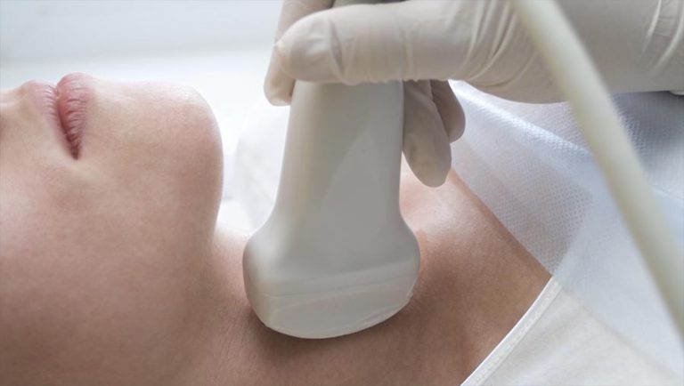
An ultrasound scan is a medical imaging examination that uses high-frequency sound waves — far above the range of human hearing — to create live, moving pictures of the inside of your body. It involves no needles, no ionising radiation and, for most examinations, no discomfort beyond gel and gentle probe pressure. It is how babies are monitored in pregnancy, how organs like the liver and kidneys are checked, how tendons and joints are assessed while they move, and how blood flow through veins and arteries is measured — all in real time, with the sonographer watching the picture form as they scan.
How does an ultrasound scan actually work?
The instrument at the heart of every scan is the transducer — the smooth handheld probe placed against your skin. It sends out pulses of sound at frequencies between roughly 1 and 20 megahertz, then listens for the echoes that bounce back as those pulses meet different tissues. Because fluid, muscle, fat and organ tissue each reflect sound differently, a computer can turn the returning echoes into a detailed greyscale image — what sonographers call B-mode imaging, the standard diagnostic picture. The gel on your skin has one simple job: sound travels poorly through air, and the gel removes the air gap so the echoes pass cleanly between probe and body.
A second mode, Doppler ultrasound, measures how sound shifts when it bounces off moving blood cells — which is how we assess flow through arteries and veins, from DVT checks to carotid artery scans. You can read more in our guide to Doppler imaging.
What is ultrasound used for?
Almost every part of the body that isn't shielded by bone or gas. In pregnancy, it confirms viability, dates the pregnancy and tracks growth. In general diagnostics, it examines the liver, gallbladder, pancreas, kidneys and bladder, and assesses lumps and swellings. Women's health scanning covers the pelvis, ovaries and breast; men's health scanning covers the testes and prostate-related symptoms. Musculoskeletal ultrasound assesses tendons, ligaments and joints — uniquely, while you move them — and vascular ultrasound maps blood flow. If you are not sure which examination fits your symptom, our Scan Advisor or a two-minute phone call will point you to the right one.
What happens at an ultrasound appointment?
Preparation depends on the scan — some abdominal examinations need a few hours of fasting, some pelvic scans need a full bladder, and many scans need nothing at all; our preparation guide covers each type. At the appointment you'll lie or sit on the examination couch, gel is applied, and the sonographer moves the transducer over the area while watching the screen — typically for 20 to 40 minutes depending on the examination. At IUS London, findings are discussed with you at the scan itself, and your written report is ready usually within two hours, written so your GP or specialist can act on it directly.
Types of ultrasound scan
Standard 2D B-mode is the diagnostic workhorse — every measurement that matters clinically is made on it. Doppler adds blood-flow information. Some examinations are performed internally for better image quality — a transvaginal scan in gynaecology, for example, brings the probe closer to the structures being examined. Dynamic scanning, used in MSK work, assesses structures while they move. You may also have heard of 3D and 4D "bonding" scans — rendered keepsake images. We do not offer these, as a deliberate clinical position: every scan must earn its place clinically.
Is ultrasound safe?
Ultrasound uses sound, not ionising radiation — there is no radiation dose, which is precisely why it is the first-line imaging of pregnancy and can be repeated as often as clinically needed. It has been used in medicine for over half a century with an excellent safety record, maintained by the prudent-use principle: scans are performed by qualified operators, at appropriate settings, for a clinical reason. We cover the evidence in detail in Is ultrasound safe?
Who performs the scan — and why it matters
Ultrasound is the most operator-dependent imaging method in medicine: the picture is only as good as the person creating and interpreting it, in the moment. At IUS London every scan is performed by an HCPC-registered sonographer with a senior NHS background — meet the team, registration numbers included. If you are comparing clinics, our guide to choosing a private ultrasound clinic explains exactly what to check.
How does ultrasound compare with X-ray, CT and MRI?
Each looks at the body differently: X-ray and CT excel at bone and use ionising radiation; MRI excels at deep soft-tissue detail; ultrasound is the real-time, radiation-free specialist of soft tissue, movement and blood flow — and usually the fastest and most accessible of the four. Our comparison guide, Ultrasound vs MRI vs CT, walks through which test suits which question.
Frequently asked questions
Does an ultrasound scan hurt?
No — you'll feel gel and gentle probe pressure. In MSK scanning the sonographer may press where it hurts or ask you to move, because reproducing your symptom under the probe is part of finding its cause.
Do I need a referral for a private ultrasound?
No — at IUS London you can self-refer for our scans, and your report is written for your GP or specialist to act on. One exception: transplant kidney evaluation requires a referral from your transplant team.
How long does a scan take?
Most examinations take 20 to 40 minutes, including time to discuss what the sonographer is seeing.
Is there any radiation?
None — ultrasound uses sound waves only, which is why it is the standard imaging of pregnancy and safe to repeat when clinically needed.
Author: Yianni Kiromitis, Senior Sonographer, HCPC RA38415 — founder of IUS London.